. 2012 Feb;40(3):1366-80.
doi: 10.1093/nar/gkr831. Epub 2011 Oct 7.

Molecular architecture of a multifunctional MCM complex

Affiliations

Molecular architecture of a multifunctional MCM complex

June Sanchez-Berrondo et al. Nucleic Acids Res. 2012 Feb.

Abstract

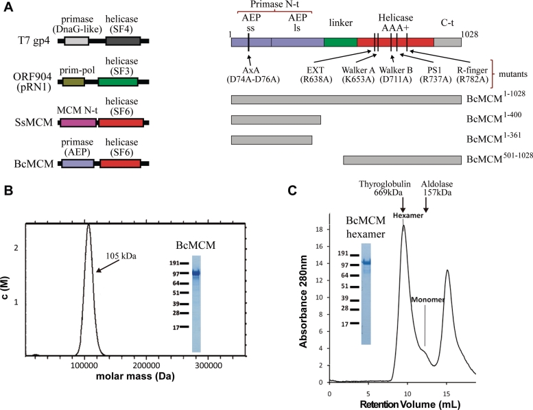
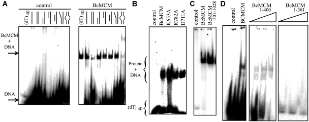
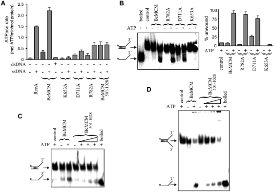
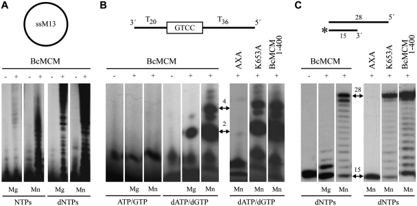
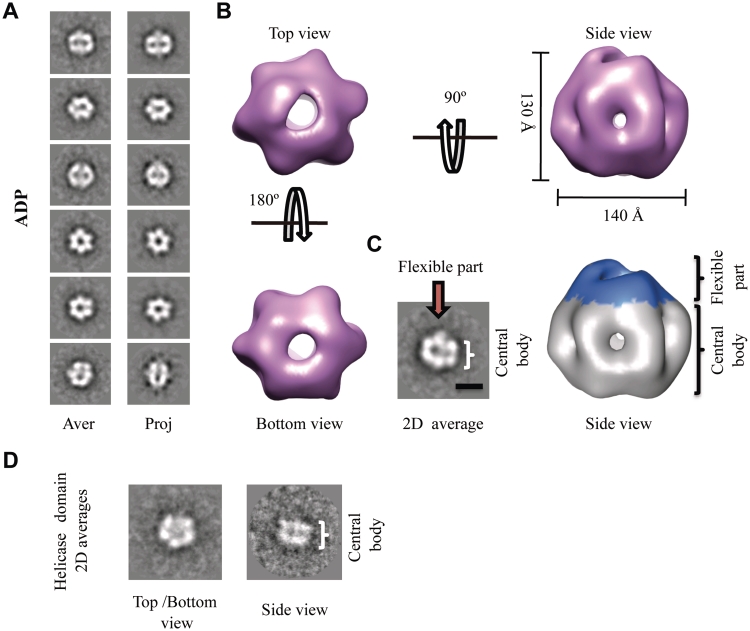
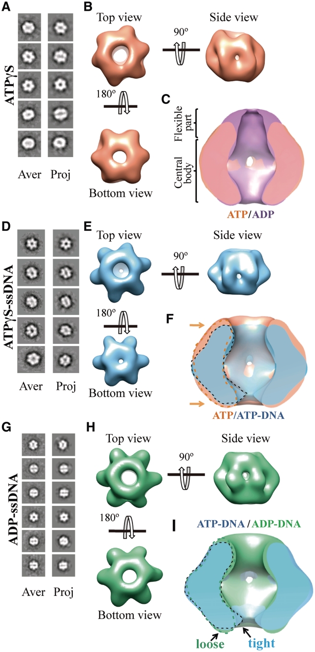
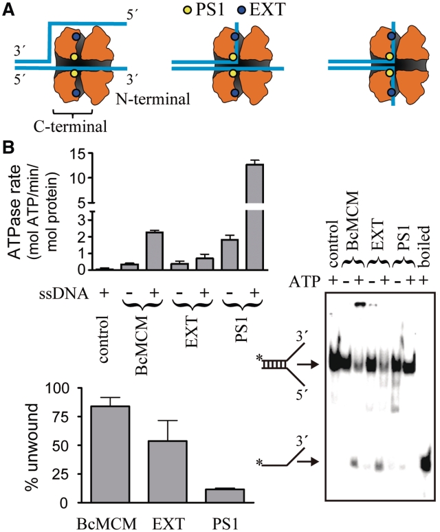
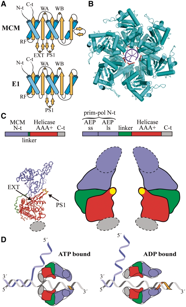
DNA replication is strictly regulated through a sequence of steps that involve many macromolecular protein complexes. One of them is the replicative helicase, which is required for initiation and elongation phases. A MCM helicase found as a prophage in the genome of Bacillus cereus is fused with a primase domain constituting an integrative arrangement of two essential activities for replication. We have isolated this helicase-primase complex (BcMCM) showing that it can bind DNA and displays not only helicase and primase but also DNA polymerase activity. Using single-particle electron microscopy and 3D reconstruction, we obtained structures of BcMCM using ATPγS or ADP in the absence and presence of DNA. The complex depicts the typical hexameric ring shape. The dissection of the unwinding mechanism using site-directed mutagenesis in the Walker A, Walker B, arginine finger and the helicase channels, suggests that the BcMCM complex unwinds DNA following the extrusion model similarly to the E1 helicase from papillomavirus.


Figures

Figure 1.
Figure 1.
BcMCM is purified as a monomer but can hexamerize. (A) Graphical representations of the domain architecture present in several multimodular helicases (left panel). The families of associated primases and helicases are indicated. BcMCM presents an exclusive combination of an archaeo–eukaryotic primase (AEP), including the small (ss) and large (ls) subunits, and a MCM-like helicase (Super Family 6). Scheme of the domain arrangement of BcMCM (right panel). A representation of the different constructions and mutants used along this article is depicted in the right lower panel. (B) Analytical ultracentrifugation experiments shows that the recombinant protein BcMCM expressed in E. coli is a monomer. Only one species is detected in the calculated molar mass distribution, c(M), which is consistent with the molecular mass of a monomer (see ‘Materials and Methods’ section). (C) Size-exclusion chromatography illustrates conversion of BcMCM monomers into hexamers. Upon addition of ATPγS and ssDNA (dT)40 and incubation with the purified monomer fractions the BcMCM protein elutes mainly as a hexamer from Superdex 200 column. The hexamer fractions were collected and analysed using SDS–PAGE (inset). The peak at the end of the chromatogram corresponds to the excess of ssDNA used in the assay. Elution positions of the molecular weight standards Thyroglobulin and Aldolase are indicated in the graph.
Figure 2.
Figure 2.
The BcMCM protein binds DNA. (A) Native EMSA assays using different DNA structures. Sketches of the different DNA probes used in the assay are indicated above each lane. The assay without BcMCM was used as negative control. The 40-nt lane corresponds to a 40-nt poly-dT, (dT)40, a probe to discard binding to DNA secondary structures, and was used in subsequent assays. The sequences of the probes are described in Supplementary Table SII. Arrows and brackets indicate positions of the shifted protein–DNA complex and free DNA. All the assays were carried out using 1 µM of protein, unless otherwise indicated, and 1 nM of DNA. (B) Mutations in the ATPase site do not affect DNA binding. (C) The helicase domain BcMCM501–1028 is able to bind DNA. (D) The canonical primase domain BcMCM1–361 (1–3 µM) does not bind DNA. However, BcMCM1–400 (1–3 µM) shows weak binding compared to the wild-type protein.
Figure 3.
Figure 3.
BcMCM hydrolyses ATP and unwinds DNA. (A) For comparison the ATP hydrolysis activity of BcMCM and the different mutants was measured and represented as ATPase rates. The presence of ssDNA stimulates BcMCM ATPase activity. However, no effect was observed in the presence of dsDNA. The mutation in the Walker A region abolishes ATP hydrolysis, whereas the mutation in Walker B or the arginine finger reduces but not abolishes ATPase activity of BcMCM. No ssDNA stimulation was observed in the case of the BcMCM501–1028. An amount of 1 µM of protein was used in each reaction. The assay was performed by triplicate and the error bars indicate SEM. (B) BcMCM unwinds DNA with 3′→5′ polarity. The experiment compares the ATP dependent ability of BcMCM protein and the different mutants to separate the strands (% unwound DNA) of a specific dsDNA. Walker B or arginine finger mutants displayed a reduced ATP hydrolysis, although still show helicase activity. However, the Walker A mutant completely abrogated DNA unwinding. The assays were carried out using 0.5 nM of dsDNA and 0.5 µM of protein. (C and D) The helicase domain BcMCM501–1028 also presents helicase activity using fork or 3′-overhang DNA substrates. The assay for BcMCM501–1028 was performed using a gradient from 0.5 to 2 µM of protein.
Figure 4.
Figure 4.
BcMCM has intrinsic primase and polymerase activity. (A) Circular single-stranded M13 was used as template for BcMCM primase activity. BcMCM primase uses dNTPs more efficiently than NTPs and the reaction is favoured in the presence of manganese as cofactor. (B) A 60-mer ssDNA oligonucleotide (see scheme) was used as template with a putative priming site at the boxed sequence. The wild-type BcMCM displayed an intrinsic primase that preferentially uses dNTPs as substrates and manganese as metal activator. The primase activity is eliminated by the double mutation AxA in the catalytic site. In the monomeric Walker A mutant (K653A) and in the truncated version BcMCM1–400 the primase activity was similar. Arrows indicate the main formation of a dinucleotide and a 4-mer products. (C) Using a conventional template/primer substrate (see scheme), BcMCM displayed an intrinsic DNA polymerization activity, which was absent in the mutant AxA. The polymerase activity is preferentially activated by manganese ions. The DNA synthesis of the hexameric BcMCM is similar to the monomeric Walker A mutant (K653A), thus DNA polymerase activity does not involve hexamerization. The DNA polymerase activity of BcMCM, as in the case of the primase activity, is normal in the truncated version BcMCM1–400. Therefore both the primase and polymerase activities do not require the helicase domain. The initial substrate and the complete product positions are depicted with two-headed arrows. The asterisk in the substrate sketch indicates the labelled primer.
Figure 5.
Figure 5.
3D reconstruction of the BcMCM–ADP hexamer. (A) Gallery of selected reference-free 2D averages (left panel) compared to the corresponding reprojections of the final structure (right panel). (B) Surface representation of the 3D reconstruction of BcMCM–ADP hexamer filtered to 36 Å shown in different orientations. The protein monomers assemble into a single hexameric ring around a large central cavity displaying differences between the top and the bottom views. The overall dimensions of the complex are depicted in the figure. (C) The BcMCM–ADP hexamer (right panel) consists of a central body indicated (coloured in grey) and a flexible apical part (coloured in blue). In the 2D average side view (left panel) the central body and the flexible part are indicated with a bracket and an arrow respectively. A 100-Å bar is provided as a reference. (D) A typical reference-free 2D class averages of the BcMCM501–1028 helicase domain resembles the 2D averages of the BcMCM complex.
Figure 6.
Figure 6.
3D reconstruction of BcMCM–ATPγS, BcMCM–ATPγS-DNA and BcMCM–ADP complexes. (A) BcMCM–ATPγS reference-free class averages (left panel) and corresponding reprojections from the final structure (right panel). (B) Several views of different surface representations of a 3D reconstruction of BcMCM–ATPγS hexamer filtered to 36 Å. (C) Superposition of the symmetrized cut-open side views of BcMCM–ADP model in magenta and the symmetrized BcMCM–ATPγS in orange. ATP hydrolysis does not introduce large conformational changes in the BcMCM central body region. (D) BcMCM–ATPγS–ssDNA reference-free class averages (left panel) and corresponding reprojections from the structure (right panel). (E) Several views of different surface representations of BcMCM–ATPγS–ssDNA hexamer filtered to 33 Å. (F) Superposition of symmetrized cut-open side views of BcMCM–ATPγS model in orange and the symmetrized BcMCM–ATPγS–ssDNA in blue. One monomer of a BcMCM–ATPγS–ssDNA is highlighted with a dashed black line while one monomer of the BcMCM–ATPγS is highlighted with a dashed orange line. Conformational changes upon DNA binding (indicated by an arrow) permit a closer interaction of the DNA with the BcMCM hexamer. (G) BcMCM–ADP–ssDNA reference-free class averages (left panel) and corresponding reprojections from the structure (right panel). (H) Different views of a surface representation of a 3D reconstruction of BcMCM–ADP–ssDNA hexamer filtered to 33 Å. (I) Superposition of symmetrized cut-open side views of BcMCM–ATPγS–ssDNA model in blue and the symmetrized BcMCM–ADP–ssDNA in green. One monomer of a BcMCM–ATPγS–ssDNA is highlighted with a black line while one monomer of the BcMCM–ADP–ssDNA is indicated with a dashed green line.
Figure 7.
Figure 7.
ATPase and helicase activity of the PS1 and the EXT mutants. (A) Schematic view of the location of the mutations in the BcMCM model and their implications in the different proposed extrusion mechanisms variants to unwind DNA (16). Each model threads the DNA strands in a different way through the helicase central and hypothetical lateral channels, which are depicted as a shaded inner surface. (B) ATPase (upper panel) and helicase (lower panel) activity of the PS1 and EXT BcMCM mutants. The histograms show the average of three experiments and the error bars indicate the SEM. A representative gel displaying the helicase experiment is depicted in the right side. Both assays were performed as in Figure 3.
Figure 8.
Figure 8.
Unwinding mechanism of BcMCM. (A) Topology diagrams of the AAA+ domain of the MCM and E1 helicases. The sketch shows the conservation of the Walker A (WA), Walker B (WB), arginine finger (RF) and PS1 motifs between both helicases. The topology models were drawn based on the SsMCM (residues 304–484) and E1 (residues 408–545) structures. (B) Structure of the E1 helicase in complex with ssDNA. The lysine residues in the PS1 motifs contacting the DNA are coloured in red. (C) Model of the domain organization in BcMCM (right panel) based on the SsMCM crystal structure (left panel) (16) and the 3D-EM structures (Figures 5, 6 and Supplementary Figures S7 and S9). AEP denotes de archaeo–eukaryotic primase domain containing the small (ss) and large (ls) subunits. The PS1 β-hairpin is depicted with a yellow oval in the sketch. The dotted lines indicate the flexible protein regions. (D) Longitudinal sections of a hypothetical model for BcMCM helicase activity based on the steric exclusion model and the active role of the PS1 β-hairpin. A dotted line derived from the EM structures encircles the domain sketches. The BcMCM hexamer would embrace the DNA using the C-terminal regions in the ATP-bound state closing the helicase side aperture. The hydrolysis of the nucleotide would trigger the reorganization of the C-terminal, widening the helicase side and facilitating the translocation of the DNA by the PS1 β-hairpin through the central channel. Concomitantly with the helicase activity the flexible primase–polymerase domain would start primer synthesis (orange fragment). Therefore a unique enzyme might unwind dsDNA, and at the same time use one of the DNA strands as a template for DNA synthesis without the need for generating RNA as an intermediate product.

References

    1. Chong JP, Hayashi MK, Simon MN, Xu RM, Stillman B. A double-hexamer archaeal minichromosome maintenance protein is an ATP-dependent DNA helicase. Proc. Natl Acad. Sci. USA. 2000;97:1530–1535. - PMC - PubMed
    1. Shechter DF, Ying CY, Gautier J. The intrinsic DNA helicase activity of Methanobacterium thermoautotrophicum delta H minichromosome maintenance protein. J. Biol. Chem. 2000;275:15049–15059. - PubMed
    1. Niedenzu T, Roleke D, Bains G, Scherzinger E, Saenger W. Crystal structure of the hexameric replicative helicase RepA of plasmid RSF1010. J. Mol. Biol. 2001;306:479–487. - PubMed
    1. Bochman ML, Schwacha A. The Mcm2-7 complex has in vitro helicase activity. Mol. Cell. 2008;31:287–293. - PubMed
    1. Gomez-Llorente Y, Fletcher RJ, Chen XS, Carazo JM, San Martin C. Polymorphism and double hexamer structure in the archaeal minichromosome maintenance (MCM) helicase from Methanobacterium thermoautotrophicum. J. Biol. Chem. 2005;280:40909–40915. - PubMed

Publication types

MeSH terms