Review
. 2012 Jan 1;302(1):H10-23.
doi: 10.1152/ajpheart.00574.2011. Epub 2011 Oct 7.

The coronary circulation in exercise training

Affiliations
Review

The coronary circulation in exercise training

M Harold Laughlin et al. Am J Physiol Heart Circ Physiol. .

Abstract

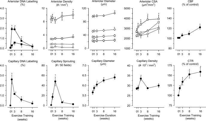
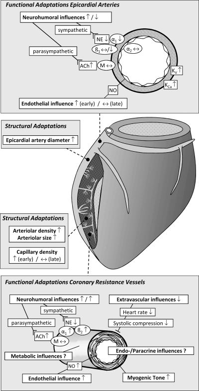
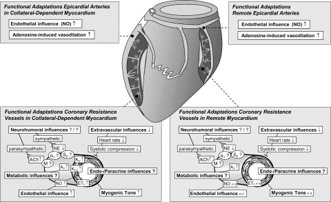
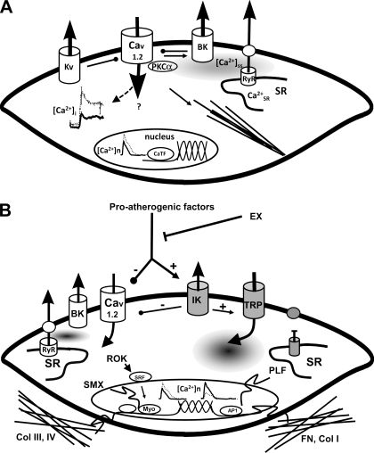
Exercise training (EX) induces increases in coronary transport capacity through adaptations in the coronary microcirculation including increased arteriolar diameters and/or densities and changes in the vasomotor reactivity of coronary resistance arteries. In large animals, EX increases capillary exchange capacity through angiogenesis of new capillaries at a rate matched to EX-induced cardiac hypertrophy so that capillary density remains normal. However, after EX coronary capillary exchange area is greater (i.e., capillary permeability surface area product is greater) at any given blood flow because of altered coronary vascular resistance and matching of exchange surface area and blood flow distribution. The improved coronary capillary blood flow distribution appears to be the result of structural changes in the coronary tree and alterations in vasoreactivity of coronary resistance arteries. EX also alters vasomotor reactivity of conduit coronary arteries in that after EX, α-adrenergic receptor responsiveness is blunted. Of interest, α- and β-adrenergic tone appears to be maintained in the coronary microcirculation in the presence of lower circulating catecholamine levels because of increased receptor responsiveness to adrenergic stimulation. EX also alters other vasomotor control processes of coronary resistance vessels. For example, coronary arterioles exhibit increased myogenic tone after EX, likely because of a calcium-dependent PKC signaling-mediated alteration in voltage-gated calcium channel activity in response to stretch. Conversely, EX augments endothelium-dependent vasodilation throughout the coronary arteriolar network and in the conduit arteries in coronary artery disease (CAD). The enhanced endothelium-dependent dilation appears to result from increased nitric oxide bioavailability because of changes in nitric oxide synthase expression/activity and decreased oxidant stress. EX also decreases extravascular compressive forces in the myocardium at rest and at comparable levels of exercise, mainly because of decreases in heart rate and duration of systole. EX does not stimulate growth of coronary collateral vessels in the normal heart. However, if exercise produces ischemia, which would be absent or minimal under resting conditions, there is evidence that collateral growth can be enhanced. While there is evidence that EX can decrease the progression of atherosclerotic lesions or even induce the regression of atherosclerotic lesions in humans, the evidence of this is not strong due to the fact that most prospective trials conducted to date have included other lifestyle changes and treatment strategies by necessity. The literature from large animal models of CAD also presents a cloudy picture concerning whether EX can induce the regression of or slow the progression of atherosclerotic lesions. Thus, while evidence from research using humans with CAD and animal models of CAD indicates that EX increases endothelium-dependent dilation throughout the coronary vascular tree, evidence that EX reverses or slows the progression of lesion development in CAD is not conclusive at this time. This suggests that the beneficial effects of EX in CAD may not be the result of direct effects on the coronary artery wall. If this suggestion is true, it is important to determine the mechanisms involved in these beneficial effects.


Figures

Fig. 1.
Fig. 1.
Effects of exercise training (EX) in swine on DNA labeling in arterioles [top, left: smooth muscle cells (■) and endothelial cells (●)], arteriolar density [top, second panel: 20–30-μm-diameter (○), 31–40-μm-diameter (□), 41–70-μm-diameter (◊), and 71–120-μm-diameter (▵) arterioles], arteriolar diameter [top, third panel: 20–30-μm-diameter (○), 31–40-μm-diameter (□), 41–70-μm-diameter (◊), and 71–120-μm-diameter (▵) arterioles], total arteriolar cross-sectional area [CSA; top, fourth panel: 20–30-μm-diameter (○), 31–40-μm-diameter (□), 41–70-μm-diameter (◊), and 71–120-μm-diameter (▵) arterioles], coronary blood flow (CBF; top, right) are shown. DNA labeling of capillaries (bottom, left), sprouting of new capillaries (bottom, second panel), capillary diameter (bottom, third panel), capillary density (bottom, fourth panel), and coronary transport reserve (CTR; bottom, right) is shown. Data are from White et al. (173), presented as means ± SE for 5 groups, with 6 animals in each group (0, 1, 3, 8 and 16 wk). *P < 0.05 different from the 0-wk time point (sedentary swine). See text for further explanation. Modified from Duncker and Bache (39) with permission from the American Physiological Society.
Fig. 2.
Fig. 2.
Summary of the structural and functional coronary microcirculatory adaptations to chronic EX in normal subjects. ACh, acetylcholine; M, muscarinic receptor; NE, norepinephrine; α1, α1-adrenergic receptor; β1, β1-adrenergic receptor; β2, β2-adrenergic receptor; Kv, voltage-gated K+ channel; KCa, Ca2+-activated K+ channel; NO, nitric oxide. Modified from Duncker and Bache (39) with permission of the American Physiological Society.
Fig. 3.
Fig. 3.
Summary of the structural and functional coronary adaptations to chronic EX in the collateral circulation. A, adenosine receptor; ET, endothelin receptor. Modified from Duncker and Bache (39) with permission of the American Physiological Society.
Fig. 4.
Fig. 4.
A: model of EX-induced adaptations of coronary smooth muscle (CSM) in normal subjects (boldface indicates those elements altered by EX). The left center illustrates decreased intracellular calcium concentration ([Ca2+]i) response to selective agonists (e.g., endothelin), which produces a reduced Ca2+-dependent activation of contraction. This decreased [Ca2+]i occurs despite an increased Ca2+-influx through L-type Ca2+ channels (Cav1.2), which is buffered by a non-sarcoplasmic reticulum (SR), non-Na+/Ca2+ exchanger mechanisms. Nuclear Ca2+ responses ([Ca2+]n) are similarly reduced by EX, which may affect Ca2+-dependent transcription factors (CaTF, e.g., cAMP response element-binding protein and nuclear factor of activated T cell) and target gene expression. On the top right note that EX increases spontaneous, slow-Ca2+ release from the SR into the subsarcolemmal space ([Ca2+]ss), which may contribute to the increased activation of large-conductance Ca2+-activated (BK) K+ channels by EX. In addition, Kv channels are also activated by EX. In arteriolar CSM, Ca2+-dependent PKC (e.g., PKCα) signaling enhances Cav1.2, leading to activation of contractile filaments and enhanced myogenic tone. Together, these changes result in an increase in the gain of the vasomotor contractile system and a more stable mature CSM phenotype. B: proposed model illustrating the manner in which EX may interact with proatherogenic factors, thereby decreasing lesion progression and/or stimulating lesion regression of atherosclerosis through regulation of CSM phenotype in coronary artery disease (CAD; gray, atherogenic effects). Proatherogenic factors (such as PDGF-BB, TNF-α, leptin) upregulate intermediate conductance Ca2+-activated K+ channels (KCa3.1, IK) and voltage-independent Ca2+ channels (e.g., TRP) while suppressing Cav1.2 and BK (KCa1.1) channels. CAD also disrupts SR Ca2+ release and extrusion and increases [Ca2+]n. As shown on the right, this ion channel profile switch enhances CSM synthesis of fibronectin (FN) and collagen I (Col I), leading to a more proinflammatory matrix composition and synthesis of proliferative genes (PLF). EX adaptations described in A produce a stable, noninflammatory phenotype and increased expression of collagens III and IV (Col III, IV), producing a noninflammatory matrix. Note that there are a number of similarities and differences in the effects of EX on CSM in normal and diseased states. RyR, ryanodine receptor; ROK, Rho-associated protein kinase; SMX, smooth muscle-specific gene expression; Myo, myocardin; AP1, activator protein-1.

References

    1. Anversa P, Levicky V, Beghi C, McDonald SL, Kikkawa Y. Morphometry of exercise-induced right ventricular hypertrophy in the rat. Circ Res 52: 57–64, 1983 - PubMed
    1. Bache RJ. Effects of hypertrophy on the cornary circulation. Prog Cardiovasc Dis 30: 403–440, 1988 - PubMed
    1. Bache RJ, Dai XZ, Schwartz JS, Homans DC. Role of adenosine in coronary vasodilation during exercise. Circ Res 62: 846–853, 1988 - PubMed
    1. Barbier J, Reland S, Ville N, Rannou-Bekono F, Wong S, Carre F. The effects of exercise training on myocardial adrenergic and muscarinic receptors. Clin Auton Res 16: 61–65, 2006 - PubMed
    1. Barnard RJ, Duncan HW, Baldwin KM, Grimditch G, Buckberg DD. Effects of intensive exercise training on myocardial performance and coronary blood flow. J Appl Physiol 49: 444–449, 1980 - PubMed

Publication types