Review
. 2012 Apr;69(7):1105-24.
doi: 10.1007/s00018-011-0847-7. Epub 2011 Oct 9.

Allosteric function and dysfunction of the prion protein

Affiliations
Review

Allosteric function and dysfunction of the prion protein

Rafael Linden et al. Cell Mol Life Sci. 2012 Apr.

Abstract

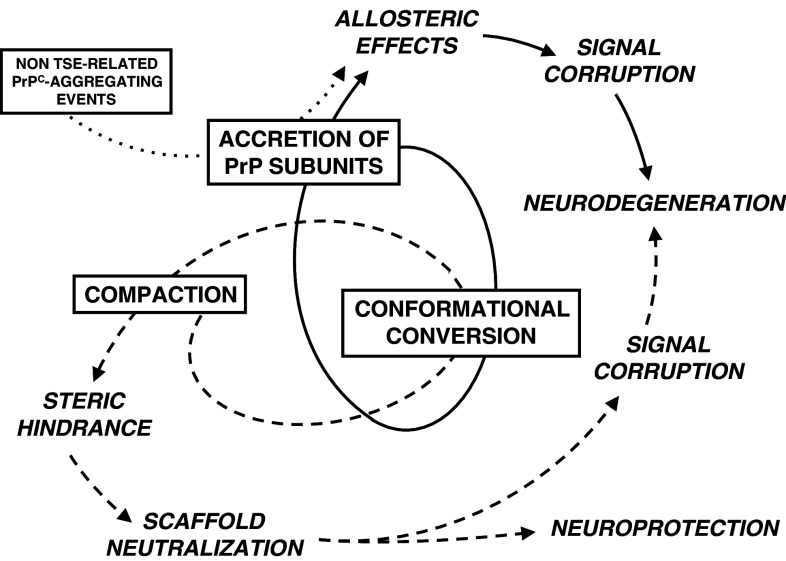
Transmissible spongiform encephalopathies (TSEs) are neurodegenerative diseases associated with progressive oligo- and multimerization of the prion protein (PrP(C)), its conformational conversion, aggregation and precipitation. We recently proposed that PrP(C) serves as a cell surface scaffold protein for a variety of signaling modules, the effects of which translate into wide-range functional consequences. Here we review evidence for allosteric functions of PrP(C), which constitute a common property of scaffold proteins. The available data suggest that allosteric effects among PrP(C) and its partners are involved in the assembly of multi-component signaling modules at the cell surface, impose upon both physiological and pathological conformational responses of PrP(C), and that allosteric dysfunction of PrP(C) has the potential to entail progressive signal corruption. These properties may be germane both to physiological roles of PrP(C), as well as to the pathogenesis of the TSEs and other degenerative/non-communicable diseases.


Figures

Fig. 1
Fig. 1
The cell surface, GPI-anchored prion protein. Drawing approximately to scale of the prion protein, showing (a) the N-terminal flexible domain; (b) location of the octapeptide repeat domain; (c) the C-terminal globular domain; (d) glycosylation chains, representing a di-glycosylated form; (e) GPI-anchor; (f) plasma membrane. Amino acid sequences are described in the legend to Fig. 4. Globular domain is depicted with Pymol software, from PDB record 1XYX of mouse PrP121-231; GPI anchor and glycosylation residues are from [142]. Modified from [239]
Fig. 2
Fig. 2
Recruitment of a PrPC-mediated signaling complex. The diagram illustrates a model of the recruitment of multicomponent cell surface complexes dependent on the presence of monomeric prion protein. a PrPC is present at the cell surface, together with nearby, non-interacting ligands, which may be either extracellular (ex) molecules (L1) or membrane proteins (L2, L3); b primary ligand L1 binds PrPC, leading to c conformational changes in either PrPC or L1, or both; d membrane partners of either PrPC (L2) or of the primary ligand L1 (L3) bind to their partners and transfer signals into the cell (in). Allosteric interactions are required to allow progression from b to c
Fig. 3
Fig. 3
Schematic diagram of the interchange between PrPC monomers and dimers. The diagram is based on PrPC crystal structures and shows hypothetical conformational transitions among two monomeric forms (A for 3HEQ.pdb and B for 1I4 M.pdb) and three possible dimers, as derived from their crystal structures. In addition to the interchain contacts stabilizing such dimers, the association between these monomers would result in the exclusion of surface area to the solvent. The α-helix of each PrPC monomer is shown in the dimers by red or cyan colors
Fig. 4
Fig. 4
Reciprocal remodeling of binding partners PrPC and hop/STI1. a Diagram of the prion protein, with indicated domains and sequences of amino acids in mouse PrPC (SP, signal peptide 1–22; OR, octapeptide repeats 51–91; CC, charged cluster 95–110; HC, hydrophobic core 112–133; H1, H2, H3, α-helices 144–153, 172–194 and 200–224, respectively; GPIp, GPI-anchoring signal 231–254), glycosylation sites (stars 180 and 196), and location of binding domains (double arrows with amino acid sequences in parentheses) of hop/STI1 (blue), neural cell adhesion molecule (N-CAM, green) and the laminin receptor precursor/laminin receptor (LRP/LR, red); b, c tri-dimensional depiction of the globular domain of PrPC, highlighting the binding domains indicated in a with the same colors; spectrometric techniques unraveled changes in alpha-helix 1 upon binding of hop/STI1230-245 to PrPC. The flexible N-terminal tail was omitted for clarity; d, e low-resolution models (sets of grey spheres) of either hop/STI1 alone (d), or the hop/STI1:PrPC complex (e), generated from SAXS measurements, reveal a compaction of the tertiary structure of hop/STI1 upon binding to PrPC (arrow points to PrPC, where the globular domain is shown in yellow and the flexible N-terminal tail is shown in pink). Modified from [34, 175, 240]
Fig. 5
Fig. 5
PrPC-dependent signaling mediated by similar allosteric effects of either hop/STI1 or the PrPC-binding peptide hop/STI1230–245: the diagrams depict a model of the behavior of PrPC, when its engagement by either the full-length hop/STI1 (a, hop) or only the peptide containing the PrPC-binding domain (b, hoppep) result in the same biological response (c, d, black arrow, see Fig. 6 a). This scenario probably requires the recruitment of a PrPC-binding, transmembrane signal transfer molecule (L2), as a consequence of allosteric effects of hop/STI1 upon PrPC (see Fig. 4b, c). Engagement of a secondary, hop/STI1-binding transmembrane protein (L3) may produce additional signals (dotted arrow) not required for the current biological response
Fig. 6
Fig. 6
Comparison of PrPC-dependent biological effects of hop/STI1 and hop/STI1230–245. a Neuroprotective effects upon undifferentiated potmitotic cells within the mouse retina. Counts of pyknotic nuclei were done in the ganglion cell layer of retinal explants cultured for 24 h with the inhibitor of protein synthesis anisomycin, either in the absence or the presence of full-length hop/STI1 or the hop/STI1 peptide containing the PrPC-binding domain (adapted from [90]). Notice the protective effects of both the full-length protein and the PrPC-binding peptide in wild-type, but not in PrPC-null nervous tissue. b Mitogenic effects on glioblastoma cell cultures. Incorporation of radioactive nucleotide was measured in cultures of the A172 glioblastoma cell line incubated for 24 h with the indicated full-length, truncated proteins or peptides (adapted from [178] and unpublished data). Notice that only the full-length protein induces cell proliferation, whereas no effect was elicited by the PrPC-binding peptide, by an irrelevant peptide nor by the truncated protein lacking the PrPC-binding domain
Fig. 7
Fig. 7
PrPC-dependent signaling mediated by a secondary ligand of hop/STI1: a, b the diagrams depict a model of the events underlying responses induced by hop/STI1, but not by hop/STI1230–245 nor by hop/STI1Δ230–245 lacking the PrPC-binding domain (Fig. 6b). The lack of response to the latter protein indicates that the response (black arrrow) depends on PrPC-hop/STI1 interaction, but the signals produced by the similar allosteric effects of either the full-length or the binding peptide (L2 and dotted arrow) upon PrPC are innefective to produce the biological response. This scenario probably requires engagement of a productive hop/STI1-binding transmembrane signal transfer molecule (L3), as a consequence of allosteric effects of PrPC upon hop/STI1 (see Fig. 4d, e)
Fig. 8
Fig. 8
Hypothetical balance of PrPC-independent and PrPC-dependent effects of a PrPC-binding protein. The diagrams depict the presumptive behavior of the PrP-binding protein hop/STI1 together with various transmembrane signal transduction molecules, in various scenarios. a hop/STI1 may bear upon various biological functions, such as cell proliferation and cell death (hatched and white arrows), independent of PrPC (see text), and signal transfer may be mediated by either one or more transmembrane molecules; b, c in certain cases, however, hop/STI1 produces conflicting signals (white vs. black arrows) via PrPC-independent and PrPC-dependent mechanisms, and the resulting biological effect may depend on stoichiometric relationships among the various components of the cell surface signaling modules; d alternative events may include intracellular networking of signaling pathways, the net result of which would also depend on the relative amounts of cell surface components
Fig. 9
Fig. 9
Multiple events of signal corruption expected from oligomerization and progressive compaction of aggregates of the prion protein. a Initial events may disrupt stoichiometrical relationships between PrPC and nearby ligands, leading to disassembly of cell surface signaling modules; b further oligo- and multimerization may lead to abnormal oligomerization of PrPC ligands, such as membrane receptors, with ensuing corruption of signal transduction through such receptors; c further compaction of aggregates of the prion protein favors steric hindrance, thus preventing binding of PrPC ligands and the assembly of cell surface signaling modules
Fig. 10
Fig. 10
Diagram of proposed pathogenic events due to dysfunction of the scaffold properties of the prion protein. The figure depicts how the asynchronous accretion of monomeric or dimeric PrPC to growing oligomers, conformational conversion of an unknown proportion of the protein molecules within agregates and progressive compaction of the aggregates may lead to the pathogenesis of TSE and other neurodegenerations. The cycle of accretion of monomers together with conformational conversion is thought to heavily impose upon the properties of PrPC-scaffolded signaling modules, leading to allosteric effects and ensuing signal corruption, which may result in neurodegeneration (arrows). Conformational conversion is, in turn, expected to fuel progressive aggregation and compaction, leading to steric hindrance, which may either cause further signal corruption and neurodegeneration, or, at late stages, contribute to protect the tissue from the recurring cycles of monomer accretion and dynamically changing allosteric effects (dashed arrows). Signal corruption due to accretion of PrPC subunits may also be triggered by non TSE-related aggregation events, such as cross-linking by Aβ oligomers (dotted line)

References

    1. Prusiner SB. Prions. Proc Natl Acad Sci USA. 1998;95(23):13363–13383. doi: 10.1073/pnas.95.23.13363. - DOI - PMC - PubMed
    1. Norrby E. Prions and protein-folding diseases. J Intern Med. 2011;270(1):1–14. doi: 10.1111/j.1365-2796.2011.02387.x. - DOI - PubMed
    1. Aguzzi A, Calella AM. Prions: protein aggregation and infectious diseases. Physiol Rev. 2009;89(4):1105–1152. doi: 10.1152/physrev.00006.2009. - DOI - PubMed
    1. Brown K, Mastrianni JA. The prion diseases. J Geriatr Psychiatry Neurol. 2010;23(4):277–298. doi: 10.1177/0891988710383576. - DOI - PubMed
    1. Caughey B, Baron GS, Chesebro B, Jeffrey M. Getting a grip on prions: oligomers, amyloids, and pathological membrane interactions. Annu Rev Biochem. 2009;78:177–204. doi: 10.1146/annurev.biochem.78.082907.145410. - DOI - PMC - PubMed

Publication types