. 2011 Oct 15;124(Pt 20):3428-40.
doi: 10.1242/jcs.083659. Epub 2011 Oct 7.

BMP2, but not BMP4, is crucial for chondrocyte proliferation and maturation during endochondral bone development

Affiliations

BMP2, but not BMP4, is crucial for chondrocyte proliferation and maturation during endochondral bone development

Bing Shu et al. J Cell Sci. .

Abstract

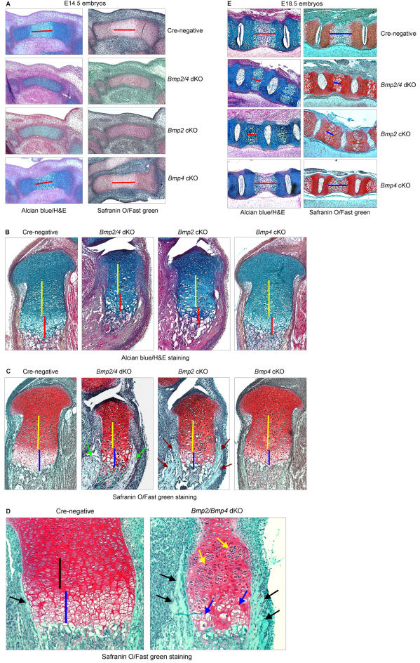
The BMP signaling pathway has a crucial role in chondrocyte proliferation and maturation during endochondral bone development. To investigate the specific function of the Bmp2 and Bmp4 genes in growth plate chondrocytes during cartilage development, we generated chondrocyte-specific Bmp2 and Bmp4 conditional knockout (cKO) mice and Bmp2,Bmp4 double knockout (dKO) mice. We found that deletion of Bmp2 and Bmp4 genes or the Bmp2 gene alone results in a severe chondrodysplasia phenotype, whereas deletion of the Bmp4 gene alone produces a minor cartilage phenotype. Both dKO and Bmp2 cKO mice exhibit severe disorganization of chondrocytes within the growth plate region and display profound defects in chondrocyte proliferation, differentiation and apoptosis. To understand the mechanism by which BMP2 regulates these processes, we explored the specific relationship between BMP2 and Runx2, a key regulator of chondrocyte differentiation. We found that BMP2 induces Runx2 expression at both the transcriptional and post-transcriptional levels. BMP2 enhances Runx2 protein levels through inhibition of CDK4 and subsequent prevention of Runx2 ubiquitylation and proteasomal degradation. Our studies provide novel insights into the genetic control and molecular mechanism of BMP signaling during cartilage development.


Figures

Fig. 1.
Fig. 1.
Skeletal growth was impaired in Bmp2/4 dKO, Bmp2 cKO, but not Bmp4 cKO embryos. (A) E18.5 Bmp2/4 dKO, Bmp2 cKO embryos and E18.0 Bmp4 cKO embryos and their littermates were collected. The whole skeleton was stained with Alizarin Red and Alcian Blue. The body size of Bmp2/4 dKO and Bmp2 cKO embryos are dramatically decreased compared with their Cre-negative littermates. (B) In Bmp2/4 dKO and Bmp2 cKO embryos heads are smaller than those of Cre-negative littermates. Cartilaginous occipital bone has almost disappeared (top panel) and the thoracic cavities and spines are much smaller than Cre-negative embryos with very little bone formation. In addition, the thoracic cavities are deformed in shape (middle panel) in these KO embryos. The hind limbs are also obviously shorter (bottom panel) in Bmp2/4 dKO and Bmp2 cKO embryos. The whole skeletal, head and hind limb of Bmp4 cKO embryos are relatively normal, with only minor defects in skeletal development (right panel).
Fig. 2.
Fig. 2.
Severe defects in growth plate cartilage morphology are found in Bmp2/4 dKO and Bmp2 cKO embryos. (A) Tibias of E14.5 Bmp2/4 dKO, Bmp2 cKO, Bmp4 cKO embryos and Cre-negative littermate controls were collected. Alcian Blue and H&E (left panel) and Safranin O and Fast Green (right panel) staining was performed in tibias sections, respectively. In Cre-negative embryos, chondrocytes in the middle of tibia begin to differentiate into hypertrophic chondrocytes and form primary ossification center, which is lightly stained with Alcian Blue or Safranin O (top panel). In Bmp2/4 dKO and Bmp2 cKO embryos, the tibia is smaller than that of Cre-negative embryo and is stained dark. There was no obvious primary ossification center formation in tibia of these KO embryos (middle panels). In Bmp4 cKO embryos, the tibia appears similar to the Cre-negative control with obvious primary ossification center formation in the middle (bottom panel). The length of the primary ossification center is marked by a red line. (BE) E18.5 tibias of Bmp2/4 dKO, Bmp2 cKO, Bmp4 cKO embryos and the Cre-negative control embryos were collected. Alcian Blue and H&E (B and E, left panel), and Safranin O and Fast green (C,D and E, right panel) staining was performed in tibia sections, respectively. Growth plate of E18.5 Cre-negative embryos shows typical columnar structure. However, growth plates of same aged Bmp2/4 dKO and Bmp2 cKO embryos are significantly smaller compared with their Cre-negative control embryos. The sizes of both proliferative zone (B,C, yellow bars; D, black bar) and hypertrophic zone (B, red bars and C, blue bars) were reduced. Disorganized chondrocytes with expanded cytoplasm and enlarged nucleus are found in hypertrophic chondrocyte area in Bmp2/4 dKO (D, blue arrows) embryos. Ectopic matrix deposition was found in the perichondrium area in Bmp2/4 dKO and Bmp2 cKO embryos (C, green and brown arrows; D, black arrows). The length of proliferative zone and hypertrophic zone of Bmp4 cKO embryos are similar to that of Cre-negative control embryos. No obvious disorganization of hypertrophic chondrocytes is found in Bmp4 cKO embryos (B,C). (E) In L4 vertebrae of E18.5 Cre-negative embryos, hypertrophic chondrocytes (red bar) occupied half of the length of vertebrae. There is vascular invasion and bone matrix formation in the middle of hypertrophic zone (top panel). Limited hypertrophic chondrocytes are found in the L4 vertebrae of Bmp2/4 dKO and Bmp2 cKO embryos (middle panel). L4 vertebrae of Bmp4 cKO embryo show similar chondrocyte hypertrophy to those of Cre-negative embryos (bottom panel).
Fig. 3.
Fig. 3.
Reduced chondrocyte proliferation and increased chondrocyte apoptosis in Bmp2/4 dKO and Bmp2 cKO embryos. (A,B) PCNA staining demonstrates that growth plate chondrocyte proliferation is reduced in E18.5 Bmp2/4 dKO and Bmp2 cKO embryos. The top panel shows corresponding histological sections stained with Alcian Blue and H&E and lower panel shows PCNA staining sections. Red arrows indicate the PCNA staining-positive cells (bottom panel). *P<0.05, unpaired Student's t-test, n=4. Values are means + s.e.m. (C) TUNEL staining demonstrates that growth plate chondrocyte apoptosis is significantly increased in E18.5 Bmp2/4 dKO and Bmp2 cKO embryos (positive-stained cells are indicated by yellow arrows). Top panel shows corresponding histological sections stained with Alcian Blue and H&E.
Fig. 4.
Fig. 4.
Chondrocyte differentiation is impaired in Bmp2/4 dKO and Bmp2 cKO embryos. (A) In situ hybridization demonstrates that Col2a1, Col10a1 and Mmp13 expression is significantly reduced in E18.5 Bmp2/4 dKO and Bmp2 cKO embryos. (BD) Total RNA was extracted from primary chondrocytes isolated from E18.5 Cre-negative control, Bmp2 cKO, Bmp4 cKO and Bmp2/4 dKO embryos. The expression of Sox9, Acan and Col2a1 genes was analyzed by real-time PCR. Results demonstrate that the expression of these chondrocyte marker genes is significantly reduced in Bmp2/4 dKO and Bmp2 cKO chondrocytes. By contrast, the expression of Sox9 and Acan was slightly but significantly reduced in Bmp4 cKO embryos. (EI) Total RNA was extracted from primary chondrocytes derived from E18.5 Bmp2 cKO and Cre-negative embryos. The expression of BMP genes was analyzed by real-time PCR. Results demonstrated that the expression of Bmp5, Bmp7, Bmp8b and Bmp9 genes was significantly reduced in Bmp2-deficient chondrocytes. (JN) Total RNA was extracted from primary chondrocytes derived from E18.5 Bmp4 cKO and Cre-negative embryos. The expression of BMP genes was analyzed by real-time PCR. Results demonstrated that the expression of Bmp5, Bmp7, Bmp8b and Bmp9 genes is not significantly changed in Bmp4-deficient chondrocytes. (O) Primary sternal chondrocytes were isolated from 3-day-old Bmp2/4fx/fx mice and were infected with Ad-Cre or Ad-GFP (control). 48 hours after infection, cells were treated with BIO (1 μM) or Wnt3a (100 ng/ml). Cell cultures were stopped 24 hours later and total RNA was extracted and expression Alp was examined by real-time PCR. BIO or Wnt3a-induced Alp upregulation is significantly inhibited in the Bmp2/4-deficient chondrocytes (Ad-Cre infected cells). *P<0.05, unpaired Student's t-test, n=3. Values are means + s.e.m.
Fig. 5.
Fig. 5.
Expression of Runx2 and Osterix is upregulated in perichondrial area of Bmp2/4 dKO and Bmp2 cKO embryos. Runx2 (A) and Osterix (B) immunohistochemistry (IHC) was performed using histological sections of E18.5 embryos. The numbers of Runx2- and Osterix-positive cells (black arrows) and staining intensity are markedly increased in perichondrial areas of Bmp2/4 dKO and Bmp2 cKO embryos. By contrast, Runx2 expression in proliferating and pre-hypertrophic areas is reduced (A).
Fig. 6.
Fig. 6.
BMP2 protects Runx2 protein degradation through inhibition of CDK4 expression. (A) CDK4 expression construct was transiently transfected into chondrogenic RCS cells. 24 hours after transfection, the cells were treated with BMP2 (100 ng/ml) for 24 hours. Runx2 and CDK4 protein expression was detected by western blotting. BMP2 inhibits CDK4 expression and enhances Runx2 protein level. Expression of CDK4 partially inhibits BMP2-induced Runx2 upregulation. (B) Runx2 ubiquitylation assay. CDK4 expression construct was transiently transfected into RCS cells. 24 hours after transfection, the cells were treated with BMP2 (100 ng/ml) for 24 hours. Proteasome inhibitor MG132 (10 μM) was added to the medium 4 hours before cell lysates were collected. Ubiquitylated proteins were pulled down using an UbiQapture-Q kit and polyubiquitylated Runx2 was detected using an anti-Runx2 antibody. BMP2 inhibits Runx2 ubiquitylation and expression of CDK4 partially reverses the inhibitory effect of BMP2 on Runx2 ubiquitylation. (C) Cdk4 siRNA was transfected into RCS cells. Cells were treated with BMP2 (100 ng/ml) with or without noggin (300 ng/ml) 24 hours after Cdk4 siRNA transfection. The expression of Runx2 and CDK4 protein was detected by western blotting. Silencing of CDK4 results in an upregulation of Runx2 protein levels. Noggin inhibits the effect of BMP2 on Runx2 upregulation but could not inhibit the effect of Cdk4 siRNA on Runx2 protein upregulation. (D) CDK4 expression construct was transiently transfected into RCS cells. Cells were treated with BMP2 for 24 hours at different concentrations (0, 20, 100, 200 ng/ml) 24 hours after CDK4 transfection. The expression of Runx2 and CDK4 protein was detected by western blotting. BMP2 upregulates Runx2 protein levels in a dose-dependent manner. Expression of CDK4 partially inhibits BMP2-induced Runx2 protein upregulation. (E) RCS cells were treated with different concentrations of BMP2 with or without transfection of CDK4. Runx2 mRNA expression was determined by real-time PCR. CDK4 partially inhibits BMP2-induced Runx2 mRNA expression. (F) RCS cells were transfected with cyclin D1 and CDK4 expression plasmids and treated with BMP2 (100 ng/ml). Cell lysates were collected and subjected to IP using an anti-cyclin-D1 or anti-CDK4 antibody followed by western blotting using the anti-CDK4 or anti-cyclin-D1 antibody. Treatment of BMP2 inhibits cyclin-D1–CDK4 interaction and addition of noggin blocks the inhibitory effect of BMP2 on cyclin-D1–CDK4 interaction.

References

    1. Akiyama H., Lyons J. P., Mori-Akiyama Y., Yang X., Zhang R., Zhang Z., Deng J. M., Taketo M. M., Nakamura T., Behringer R. R., et al. (2004). Interactions between Sox9 and beta-catenin control chondrocyte differentiation. Genes. Dev. 18, 1072-1087 - PMC - PubMed
    1. Alden T. D., Pittman D. D., Hankins G. R., Beres E. J., Engh J. A., Das S., Hudson S. B., Kerns K. M., Kallmes D. F., Helm G. A. (1999). In vivo endochondral bone formation using a bone morphogenetic protein 2 adenoviral vector. Human Gene. Ther. 10, 2245-2253 - PubMed
    1. Bandyopadhyay A., Tsuji K., Cox K., Harfe B. D., Rosen V., Tabin C. J. (2006). Genetic analysis of the roles of BMP2, BMP4, and BMP7 in limb patterning and skeletogenesis. PLoS Genet. 21, e216 - PMC - PubMed
    1. Chen D., Harris M. A., Rossini G., Dunstan C. R., Dallas S. L., Feng J. Q., Mundy G. R., Harris S. E. (1997). Bone morphogenetic protein 2 (BMP-2) enhances BMP-3, BMP-4 and bone cell differentiation marker gene expression during the induction of mineralized bone matrix formation in cultures of fetal rat calvarial osteoblasts. Calcif. Tissue Int. 60, 283-290 - PubMed
    1. Chen D., Ji X., Harris M. A., Feng J. Q., Celeste A. J., Rosen V., Mundy G. R., Harris S. E. (1998). Differential roles for BMP receptor type IB and IA in differentiation and specification of mesenchymal precursor cells to osteoblast and adipocyte lineages. J. Cell Biol. 142, 295-305 - PMC - PubMed

Publication types

MeSH terms

Substances