. 2011 Dec 2;286(48):41312-41322.
doi: 10.1074/jbc.M111.290973. Epub 2011 Oct 7.

ATP synthase complex of Plasmodium falciparum: dimeric assembly in mitochondrial membranes and resistance to genetic disruption

Affiliations

ATP synthase complex of Plasmodium falciparum: dimeric assembly in mitochondrial membranes and resistance to genetic disruption

Praveen Balabaskaran Nina et al. J Biol Chem. .

Abstract

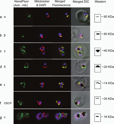
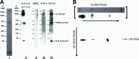
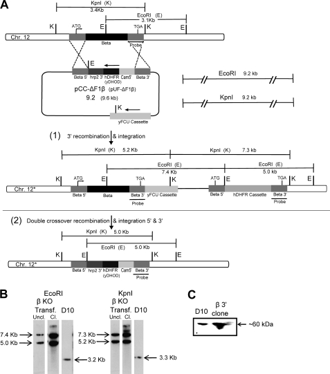
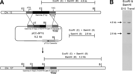
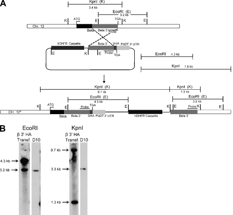
The rotary nanomotor ATP synthase is a central player in the bioenergetics of most organisms. Yet the role of ATP synthase in malaria parasites has remained unclear, as blood stages of Plasmodium falciparum appear to derive ATP largely through glycolysis. Also, genes for essential subunits of the F(O) sector of the complex could not be detected in the parasite genomes. Here, we have used molecular genetic and immunological tools to investigate the localization, complex formation, and functional significance of predicted ATP synthase subunits in P. falciparum. We generated transgenic P. falciparum lines expressing seven epitope-tagged canonical ATP synthase subunits, revealing localization of all but one of the subunits to the mitochondrion. Blue native gel electrophoresis of P. falciparum mitochondrial membranes suggested the molecular mass of the ATP synthase complex to be greater than 1 million daltons. This size is consistent with the complex being assembled as a dimer in a manner similar to the complexes observed in other eukaryotic organisms. This observation also suggests the presence of previously unknown subunits in addition to the canonical subunits in P. falciparum ATP synthase complex. Our attempts to disrupt genes encoding β and γ subunits were unsuccessful, suggesting an essential role played by the ATP synthase complex in blood stages of P. falciparum. These studies suggest that, despite some unconventional features and its minimal contribution to ATP synthesis, P. falciparum ATP synthase is localized to the parasite mitochondrion, assembled as a large dimeric complex, and is likely essential for parasite survival.


Figures

FIGURE 1.
FIGURE 1.
Localization of epitope-tagged ATP synthase subunits detected using immunofluorescence microscopy. Immunofluorescence of α (a), γ (c), δ (d), ϵ (e), OSCP (f), and c (g) subunits expressed under control of the mRPL2 promoter (see under “Experimental Procedures”). Expression of the β subunit (b) was under the control of its own promoter (see under “Experimental Procedures” and Fig. 5). Green fluorescence, anti-HA antibody (AlexaFluor 488-conjugated secondary antibody). Red fluorescence, Mitotracker Red CM-H2XRos. Blue fluorescence, DAPI (4′-6-diamidino-2-phenylindole). DIC, differential interference contrast microscopy.
FIGURE 2.
FIGURE 2.
BN-PAGE analysis of complex formation. A, P. falciparum mitochondria solubilized with digitonin were run on a BN-polyacrylamide gel (4–16% gradient) and stained with colloidal Coomassie Blue (strip I); a two-lane strip from a BN gel that was run with mitochondria isolated from β-3×HA transgenic parasites and with mitochondria isolated from parental D10 parasites and probed with monoclonal anti-HA antibody (strip II); BN gel strips run with mitochondria from β-3×HA parasites and probed with polyclonal γ and β antisera, respectively (strips III and IV); BN strip probed with normal mouse serum (strip 0, NMS). Digitonin was used at a concentration of 5 μg of detergent/μg of protein, and ∼30–40 μg of protein was loaded in each lane. Strips II–IV are blots from separate BN-polyacrylamide gels, but comparison of marker lanes indicated that the runs and transfers were highly reproducible, and they have the same marker protein size scales, within the uncertainty of the technique. B, strip from a one-dimensional (1D) BN gel was placed on top of a 4–12% SDS-polyacrylamide gel and was run in the second dimension. The SDS gel was then blotted onto a PVDF membrane and was probed with monoclonal anti-HA antibody. The gel lane and anti-HA blot strip shown above the two-dimensional (2D) blot are reproduced from A (strips I and II) to indicate the approximate position of protein (which is obscured by excess dye in the original unstained BN polyacrylamide gels) and of anti-HA immunoreactive protein in the first dimension.
FIGURE 3.
FIGURE 3.
ATP synthase β subunit gene could not be disrupted. A, diagram of the β gene locus and the vectors used for gene disruption by double crossover recombination at the β gene. In the disruption constructs, the hDHFR/yDHOD cassette was flanked by 5′ and 3′ inserts of the β gene (see under “Experimental Procedures”); the plasmids also contain the negative selectable marker yFCU. With pCC-ΔF1β, 3′ integration at the β gene (diagrammed in A(1)) was observed (B); however, no double crossover replacement (A(2)) was observed with either knock-out construct. B, Southern blot analysis of genomic DNA from transfected parasites digested separately with EcoRI and KpnI and using the probe indicated in A shows 3′ integration in uncloned (Uncl.) and cloned (CL) β KO transgenic parasites (EcoRI, 7.4, 5.0 kb; KpnI, 7.3, and 5.2 kb). Southern blot analysis of D10 genomic DNA cut with EcoRI and KpnI results in 3.2- and 3.3-kb bands, respectively. White spaces between the lanes in B indicate the cropping of irrelevant and unused lanes for clarity. C, Western blot of parasite lysates of D10 and a β KO 3′ integrant clone probed with β polyclonal antibody confirms expression of the β subunit protein.
FIGURE 4.
FIGURE 4.
ATP synthase γ subunit gene could not be disrupted. A, schematic of the knock-out strategy utilized for the attempted disruption of the γ gene. B, Southern blot of DNA recovered from transfected parasites after three cycles of selection (see under “Experimental Procedures”) and digested with EcoRI and BamHI shows a 4.8-kb fragment, as in the DNA from wild type D10 parasites, and a 2.6-kb band indicative of the continued presence of the episomal γ construct.
FIGURE 5.
FIGURE 5.
Integration of an epitope tag sequence at the end of the β gene, indicating locus accessibility. A, schematic of the β gene locus and the vector used for the integration of a 3×HA tag sequence at the 3′ end of the β gene by allelic exchange. B, Southern blot analysis of genomic DNA from transfected parasites digested with EcoRI/KpnI shows 3′ integration in β 3′ HA transgenic parasites (EcoRI, 4.3 and 3.2 kb; KpnI, 9.7 and 1.3 kb). A Southern blot of D10 wild type genomic DNA with EcoRI and KpnI results in 3.2- and 3.3-kb bands, respectively. Expression of the tagged protein is shown in Fig. 1b.

References

    1. Walker J. E., Dickson V. K. (2006) Biochim. Biophys. Acta 1757, 286–296 - PubMed
    1. Devenish R. J., Prescott M., Rodgers A. J. (2008) Int. Rev. Cell Mol. Biol. 267, 1–58 - PubMed
    1. Angevine C. M., Herold K. A., Vincent O. D., Fillingame R. H. (2007) J. Biol. Chem. 282, 9001–9007 - PubMed
    1. Fry M., Beesley J. E. (1991) Parasitology 102, 17–26 - PubMed
    1. Krungkrai J., Burat D., Kudan S., Krungkrai S., Prapunwattana P. (1999) Southeast Asian J. Trop. Med. Public Health 30, 636–642 - PubMed

Publication types

MeSH terms