

Association of calcineurin with the COPI protein Sec28 and the COPII protein Sec13 revealed by quantitative proteomics
- PMID: 21984910
- PMCID: PMC3184950
- DOI: 10.1371/journal.pone.0025280
Association of calcineurin with the COPI protein Sec28 and the COPII protein Sec13 revealed by quantitative proteomics
Abstract
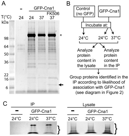
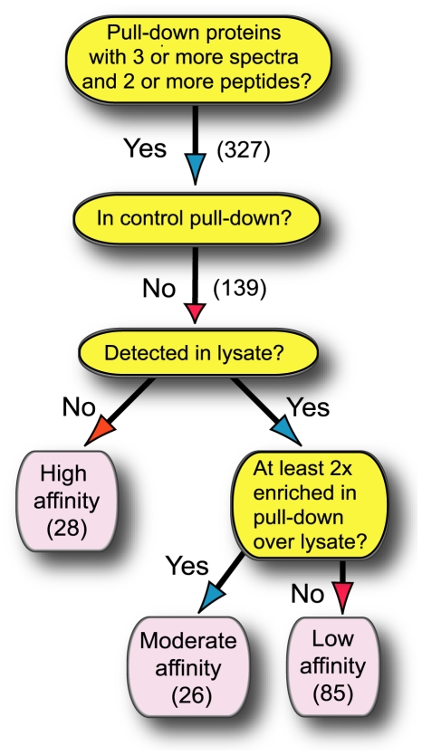
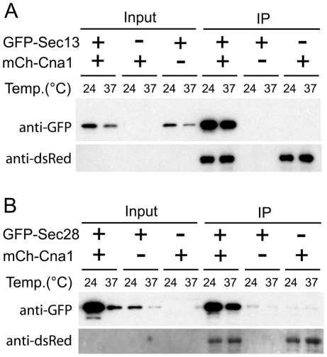
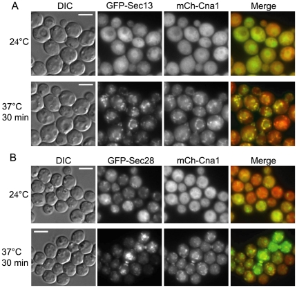
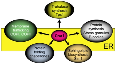
Calcineurin is a calcium-calmodulin-dependent serine/threonine specific protein phosphatase operating in key cellular processes governing responses to extracellular cues. Calcineurin is essential for growth at high temperature and virulence of the human fungal pathogen Cryptococcus neoformans but the underlying mechanism is unknown. We performed a mass spectrometry analysis to identify proteins that associate with the calcineurin A catalytic subunit (Cna1) in C. neoformans cells grown under non-stress and high temperature stress conditions. A novel prioritization strategy for mass spectrometry data from immunoprecipitation experiments identified putative substrates and proteins potentially operating with calcineurin in common pathways. Cna1 co-purified with proteins involved in membrane trafficking including the COPI component Sec28 and the COPII component Sec13. The association of Cna1 with Sec28 and Sec13 was confirmed by co-immunoprecipitation. Cna1 exhibited a dramatic change in subcellular localization during high temperature stress from diffuse cytoplasmic to ER-associated puncta and the mother-bud neck and co-localized with Sec28 and Sec13.
Conflict of interest statement
Figures
Comment in
-
A proteomic interrogation of Cryptococcus neoformans: interaction networks for calcineurin in a heated environment.Expert Rev Proteomics. 2012;9(1):13-5. doi: 10.1586/epr.11.76. Expert Rev Proteomics. 2012. PMID: 22292819
Similar articles
-
The C2 domain protein Cts1 functions in the calcineurin signaling circuit during high-temperature stress responses in Cryptococcus neoformans.Eukaryot Cell. 2011 Dec;10(12):1714-23. doi: 10.1128/EC.05148-11. Epub 2011 Oct 14. Eukaryot Cell. 2011. PMID: 22002655 Free PMC article.
-
Calcineurin colocalizes with P-bodies and stress granules during thermal stress in Cryptococcus neoformans.Eukaryot Cell. 2011 Nov;10(11):1396-402. doi: 10.1128/EC.05087-11. Epub 2011 Jul 1. Eukaryot Cell. 2011. PMID: 21724937 Free PMC article.
-
A proteomic interrogation of Cryptococcus neoformans: interaction networks for calcineurin in a heated environment.Expert Rev Proteomics. 2012;9(1):13-5. doi: 10.1586/epr.11.76. Expert Rev Proteomics. 2012. PMID: 22292819
-
Coping with stress: calmodulin and calcineurin in model and pathogenic fungi.Biochem Biophys Res Commun. 2003 Nov 28;311(4):1151-7. doi: 10.1016/s0006-291x(03)01528-6. Biochem Biophys Res Commun. 2003. PMID: 14623301 Review.
-
All about that fat: Lipid modification of proteins in Cryptococcus neoformans.J Microbiol. 2016 Mar;54(3):212-22. doi: 10.1007/s12275-016-5626-6. Epub 2016 Feb 27. J Microbiol. 2016. PMID: 26920881 Free PMC article. Review.
Cited by
-
Depletion of ε-COP in the COPI Vesicular Coat Reduces Cleistothecium Production in Aspergillus nidulans.Mycobiology. 2015 Mar;43(1):31-6. doi: 10.5941/MYCO.2015.43.1.31. Epub 2015 Mar 31. Mycobiology. 2015. PMID: 25892912 Free PMC article.
-
Calcineurin Targets Involved in Stress Survival and Fungal Virulence.PLoS Pathog. 2016 Sep 9;12(9):e1005873. doi: 10.1371/journal.ppat.1005873. eCollection 2016 Sep. PLoS Pathog. 2016. PMID: 27611567 Free PMC article.
-
Calcineurin in fungal virulence and drug resistance: Prospects for harnessing targeted inhibition of calcineurin for an antifungal therapeutic approach.Virulence. 2017 Feb 17;8(2):186-197. doi: 10.1080/21505594.2016.1201250. Epub 2016 Jun 20. Virulence. 2017. PMID: 27325145 Free PMC article. Review.
-
Estrogen receptor antagonists are anti-cryptococcal agents that directly bind EF hand proteins and synergize with fluconazole in vivo.mBio. 2014 Feb 11;5(1):e00765-13. doi: 10.1128/mBio.00765-13. mBio. 2014. PMID: 24520056 Free PMC article.
-
Calcineurin as a Multifunctional Regulator: Unraveling Novel Functions in Fungal Stress Responses, Hyphal Growth, Drug Resistance, and Pathogenesis.Fungal Biol Rev. 2014 Oct;28(2-3):56-69. doi: 10.1016/j.fbr.2014.02.004. Fungal Biol Rev. 2014. PMID: 25383089 Free PMC article.
References
-
- Cyert MS. Calcineurin signaling in Saccharomyces cerevisiae: how yeast go crazy in response to stress. Biochem Biophys Res Commun. 2003;311:1143–1150. - PubMed
-
- Crabtree GR, Olson EN. NFAT signaling: choreographing the social lives of cells. Cell. 2002;109(Suppl):S67–79. - PubMed
-
- Liu JP, Sim AT, Robinson PJ. Calcineurin inhibition of dynamin I GTPase activity coupled to nerve terminal depolarization. Science. 1994;265:970–973. - PubMed
Publication types
MeSH terms
Substances
Grants and funding
LinkOut - more resources
Full Text Sources
