. 2011;6(10):e25557.
doi: 10.1371/journal.pone.0025557. Epub 2011 Oct 3.

Contribution of the lipopolysaccharide to resistance of Shigella flexneri 2a to extreme acidity

Affiliations

Contribution of the lipopolysaccharide to resistance of Shigella flexneri 2a to extreme acidity

Mara Martinić et al. PLoS One. 2011.

Abstract

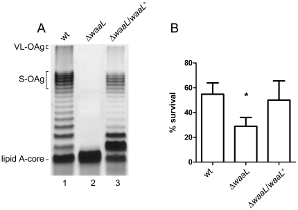
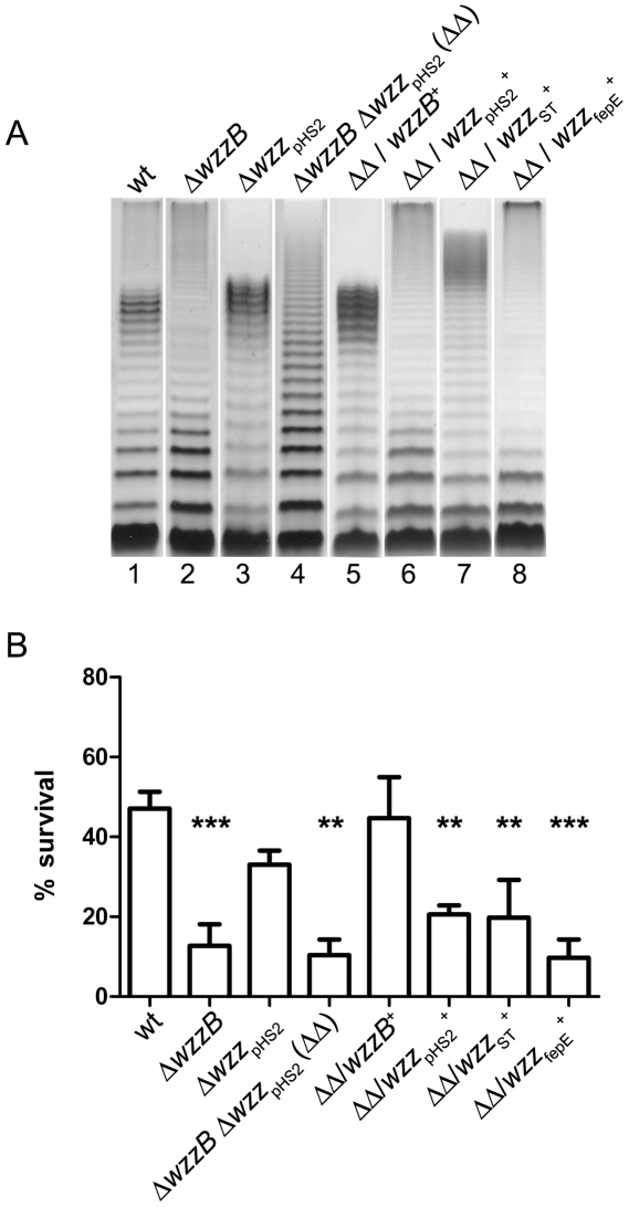
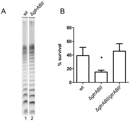
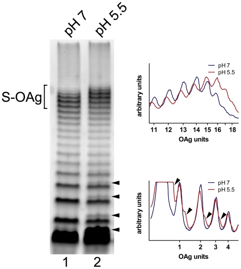
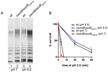
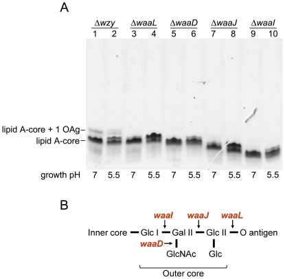
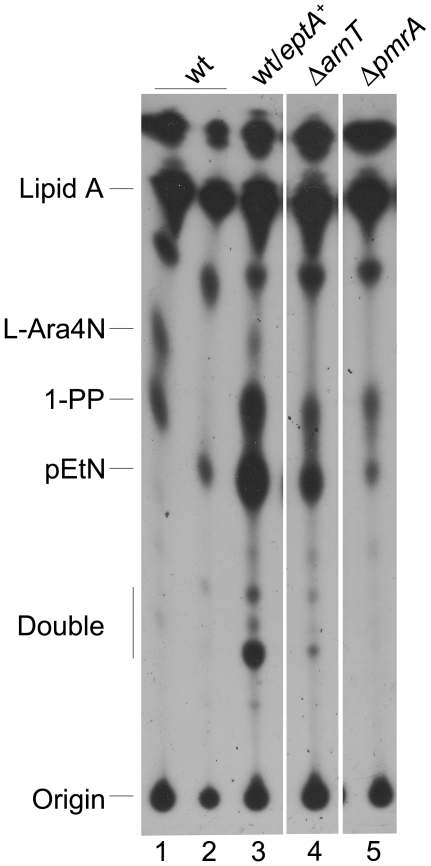
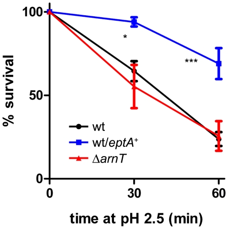
Shigella flexneri is endemic in most underdeveloped countries, causing diarrheal disease and dysentery among young children. In order to reach its target site, the colon, Shigella must overcome the acid environment of the stomach. Shigella is able to persist in this stressful environment and, because of this ability it can initiate infection following the ingestion of very small inocula. Thus, acid resistance is considered an important virulence trait of this bacterium. It has been reported that moderate acid conditions regulate the expression of numerous components of the bacterial envelope. Because the lipopolysaccharide (LPS) is the major component of the bacterial surface, here we have addressed the role of LPS in acid resistance of S. flexneri 2a. Defined deletion mutants in genes encoding proteins involved in the synthesis, assembly and length regulation of the LPS O antigen were constructed and assayed for resistance to pH 2.5 after adaptation to pH 5.5. The results showed that a mutant lacking O antigen was significantly more sensitive to extreme acid conditions than the wild type. Not only the presence of polymerized O antigen, but also a particular polymer length (S-OAg) was required for acid resistance. Glucosylation of the O antigen also contributed to this property. In addition, a moderate acidic pH induced changes in the composition of the lipid A domain of LPS. The main modification was the addition of phosphoethanolamine to the 1' phosphate of lipid A. This modification increased resistance of S. flexneri to extreme acid conditions, provide that O antigen was produced. Overall, the results of this work point out to an important role of LPS in resistance of Shigella flexneri to acid stress.


Conflict of interest statement

Competing Interests: The authors have declared that no competing interests exist.

Figures

Figure 1
Figure 1. Contribution of the O antigen to S. flexneri 2a acid resistance.
LPS profiles (A) and acid resistance (B) of S. flexneri 2457T (wt), MSF1210 (ΔwaaL) and MSF1210/pMM112 (ΔwaaL/waaL +). LPS samples from equal numbers of bacterial cells (1×10⁷ CFU) were loaded in each lane and were analyzed by Tricine-SDS-polyacrylamide gel electrophoresis on a 14% (w/v) acrylamide gel followed by silver staining. Brackets indicate the VL-OAg, the S-OAg and the lipid A-core region. For acid resistance assays, cells were grown overnight in citrate-buffered LB (pH 5.5) and diluted 1∶1000 into the acid-challenge media. Survival is stated as a percentage of the inoculum. Averages±standard errors (error bars) are shown. Statistical significance was determined by a Student's t test. (*, P <0.05).
Figure 2
Figure 2. The S. flexneri S-OAg is required for acid resistance.
LPS profiles (A) and acid resistance (B) of S. flexneri 2457T, mutants in the chain length regulators and mutants complemented with homologous or heterologous chain length regulators. Strains are 2457T (wt), MSF102 (ΔwzzB), MSF107 (Δwzz pHS2::aph), MSF209 (ΔwzzB Δwzz pHS2::aph), MSF209/pJC139, MSF209/pJC144, MSF209/pMM110 and MSF209/pJC142. LPS samples from equal numbers of bacterial cells (1×10⁷ CFU) were loaded in each lane and were analyzed by Tricine-SDS-polyacrylamide gel electrophoresis on a 14% (w/v) acrylamide gel followed by silver staining. For acid resistance assays, cells were grown overnight in citrate-buffered LB (pH 5.5) and diluted 1∶1000 into the acid-challenge media. Survival is stated as a percentage of the inoculum. Averages±standard errors (error bars) are shown. Statistical significance was determined by a Student's t test. (**, P <0.01, ***, P <0.001).
Figure 3
Figure 3. Glucosylation of the O antigen contributes to acid resistance.
(A) LPS profiles of S. flexneri 2457T (wt) and MSF2743 (ΔgtrABII). LPS samples from equal numbers of bacterial cells (1×10⁷ CFU) were loaded in each lane and were analyzed by Tricine-SDS-polyacrylamide gel electrophoresis on a 14% (w/v) acrylamide gel followed by silver staining. (B) Acid resistance of S. flexneri 2457T (wt), MSF2743 (ΔgtrABII) and MSF2743/pMM111 (ΔgtrABII/gtrABII +). Cells were grown overnight in citrate-buffered LB (pH 5.5) and diluted 1∶1000 into the acid-challenge media. Survival is stated as a percentage of the inoculum. Averages±standard errors (error bars) are shown. Statistical significance was determined by a Student's t test. (*, P <0.05).
Figure 4
Figure 4. Effect of pH on LPS profiles from S. flexneri 2457T.
Cells were grown at pH 7.0 or 5.5 and LPS samples were obtained. LPS samples from equal numbers of bacterial cells (1×10⁷ CFU) were loaded in each lane and were analyzed by Tricine-SDS-polyacrylamide gel electrophoresis on a 14% (w/v) acrylamide gel followed by silver staining. Bracket shows the S-OAg region. Arrow heads point to the double bands observed in LPS from bacteria grown at pH 5.5. The right panels show the densitograms of the bands in the gel. Upper graph shows the bands corresponding to the S-OAg (11 to 18 units) and lower graph shows the bands corresponding to the Lipid A-core substituted with 1 to 4 units.
Figure 5
Figure 5. Changes in the electrophoretic mobility of the LPS induced by moderate acidic conditions are not relevant to resistance to extreme acid.
LPS profiles (A) and acid resistance (B) of S. flexneri 2457T (wt) and MSF102 (ΔwzzB) transformed with plasmids pRMCD108 (WzzB(K267N)) or pRMCD127 (WzzB(M32T)). Bacteria were grown at pH 7.0 or 5.5 and LPS samples were obtained. LPS samples from equal numbers of bacterial cells (1×10⁷ CFU) were loaded in each lane and were analyzed by Tricine-SDS-polyacrylamide gel electrophoresis on a 14% (w/v) acrylamide gel followed by silver staining. For acid resistance assays, cells were grown overnight in citrate-buffered LB (pH 5.5) and diluted 1∶1000 into the acid-challenge media. Survival is stated as a percentage of counts at time zero. Averages±standard errors (error bars) are shown. Statistical significance was determined by the two-way ANOVA and Bonferroni post test. No significant differences were found in bacteria previously grown at pH 5.5 (closed symbols) or pH 7.0 (open symbols).
Figure 6
Figure 6. LPS profiles from S. flexneri mutants defective in O antigen and core synthesis and proposed structure of the outer core of S. flexneri 2a.
(A) Bacteria were grown at pH 7.0 or 5.5. LPS samples from equal numbers of bacterial cells (1×10⁷ CFU) were loaded in each lane and were analyzed by Tricine-SDS-polyacrylamide gel electrophoresis on a 14% (w/v) acrylamide gel followed by silver staining. The strains are MSF1749 (Δwzy), MSF1210 (ΔwaaL), MSF1144 (ΔwaaD), MSF1009 (ΔwaaI) and MSF1015 (ΔwaaJ). (B) Genes encoding proteins involved in the addition of the different sugars during outer core biosynthesis are indicated in red.
Figure 7
Figure 7. Effect of pH on lipid A modifications in S. flexneri.
32P-labelled lipid A was isolated from bacteria grown in N-minimal medium at pH 7.0 (lanes 1, 3, 4 and 5) or 5.5 (lane 2). Lipid A species were resolved by TLC with the solvent system chloroform/pyridine/88% formic acid/water (50∶50∶16∶5, v/v). The strains are 2457T (wt), 2457T/pMM113 (wt/eptA +), MSF1650 (ΔarnT) and MSF1666 (ΔpmrA).
Figure 8
Figure 8. Effect of lipid A modifications on acid resistance of S. flexneri.
The strain are 2457T (wt), 2457T/pMM113 (wt/eptA +) and MSF1650 (ΔarnT). Cells were grown overnight in citrate-buffered LB (pH 5.5) and diluted 1∶1000 into the acid-challenge media. Survival is stated as a percentage of counts at time zero. Averages±standard errors (error bars) are shown. Statistical significance was determined by. the two-way ANOVA and Bonferroni post test (*, P <0.05, ***, P <0.001).

References

    1. Kotloff KL, Winickoff JP, Ivanoff B, Clemens JD, Swerdlow D, et al. Global burden of Shigella infections: implications for vaccine development and implementation of control strategies. Bull W H O. 1999;77:651–666. - PMC - PubMed
    1. Schroeder GN, Hilbi H. Molecular pathogenesis of Shigella spp.: controlling host cell signaling, invasion, and death by type III secretion. Clin Microbiol Rev. 2008;21:134–156. - PMC - PubMed
    1. Blaser MJ, Newman LS. A review of human salmonellosis: I. Infective dose. Rev Infect Dis. 1982;4:1096–1106. - PubMed
    1. Gorden J, Small PLC. Acid resistance in enteric bacteria. Infect Immun. 1993;61:364–367. - PMC - PubMed
    1. Audia JP, Webb CC, Foster JW. Breaking through the acid barrier: an orchestrated response to proton stress by enteric bacteria. Int J Med Microbiol. 2001;29:97–106. - PubMed

Publication types

MeSH terms