

Micro-CT based experimental liver imaging using a nanoparticulate contrast agent: a longitudinal study in mice
- PMID: 21984939
- PMCID: PMC3184160
- DOI: 10.1371/journal.pone.0025692
Micro-CT based experimental liver imaging using a nanoparticulate contrast agent: a longitudinal study in mice
Abstract
Background: Micro-CT imaging of liver disease in mice relies on high soft tissue contrast to detect small lesions like liver metastases. Purpose of this study was to characterize the localization and time course of contrast enhancement of a nanoparticular alkaline earth metal-based contrast agent (VISCOVER ExiTron nano) developed for small animal liver CT imaging.
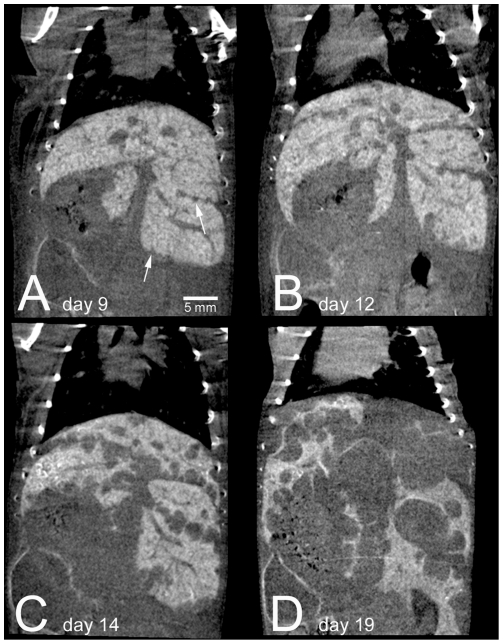
Methodology: ExiTron nano 6000 and ExiTron nano 12000, formulated for liver/spleen imaging and angiography, respectively, were intravenously injected in C57BL/6J-mice. The distribution and time course of contrast enhancement were analysed by repeated micro-CT up to 6 months. Finally, mice developing liver metastases after intrasplenic injection of colon carcinoma cells underwent longitudinal micro-CT imaging after a single injection of ExiTron nano.
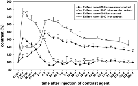
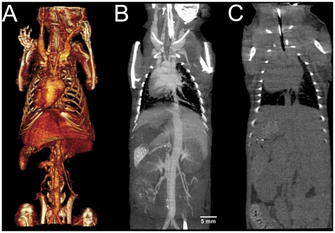
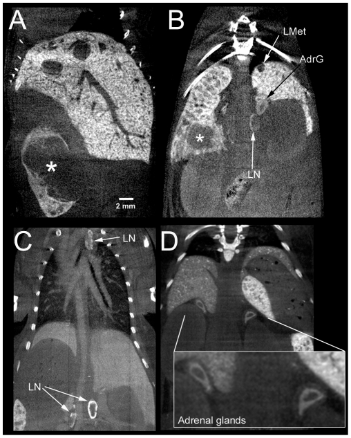
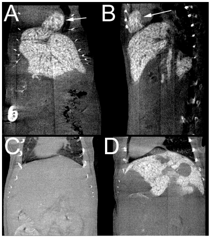
Principal findings: After a single injection of ExiTron nano the contrast of liver and spleen peaked after 4-8 hours, lasted up to several months and was tolerated well by all mice. In addition, strong contrast enhancement of abdominal and mediastinal lymph nodes and the adrenal glands was observed. Within the first two hours after injection, particularly ExiTron nano 12000 provided pronounced contrast for imaging of vascular structures. ExiTron nano facilitated detection of liver metastases and provided sufficient contrast for longitudinal observation of tumor development over weeks.
Conclusions: The nanoparticulate contrast agents ExiTron nano 6000 and 12000 provide strong contrast of the liver, spleen, lymph nodes and adrenal glands up to weeks, hereby allowing longitudinal monitoring of pathological processes of these organs in small animals, with ExiTron nano 12000 being particularly optimized for angiography due to its very high initial vessel contrast.
Conflict of interest statement
Figures
References
-
- Schambach SJ, Bag S, Schilling L, Groden C, Brockmann MA. Application of micro-CT in small animal imaging. Methods. 2010;50:2–13. - PubMed
-
- Willekens I, Lahoutte T, Buls N, Vanhove C, Deklerck R, et al. Time-course of contrast enhancement in spleen and liver with Exia 160, Fenestra LC, and VC. Mol Imaging Biol. 2009;11:128–135. - PubMed
-
- Suckow CE, Stout DB. MicroCT liver contrast agent enhancement over time, dose, and mouse strain. Mol Imaging Biol. 2008;10:114–120. - PubMed
-
- Graham KC, Detombe SA, MacKenzie LT, Holdsworth DW, MacDonald IC, et al. Contrast-enhanced microcomputed tomography using intraperitoneal contrast injection for the assessment of tumor-burden in liver metastasis models. Invest Radiol. 2008;43:488–495. - PubMed
-
- Montet X, Pastor CM, Vallee JP, Becker CD, Geissbuhler A, et al. Improved visualization of vessels and hepatic tumors by micro-computed tomography (CT) using iodinated liposomes. Invest Radiol. 2007;42:652–658. - PubMed
Publication types
MeSH terms
Substances
LinkOut - more resources
Full Text Sources
