. 2011 Jul;1(2):170-85.
doi: 10.1158/2159-8290.CD-11-0039. Epub 2011 Jun 7.

High frequency of PIK3R1 and PIK3R2 mutations in endometrial cancer elucidates a novel mechanism for regulation of PTEN protein stability

Affiliations

High frequency of PIK3R1 and PIK3R2 mutations in endometrial cancer elucidates a novel mechanism for regulation of PTEN protein stability

Lydia W T Cheung et al. Cancer Discov. 2011 Jul.

Erratum in

Abstract

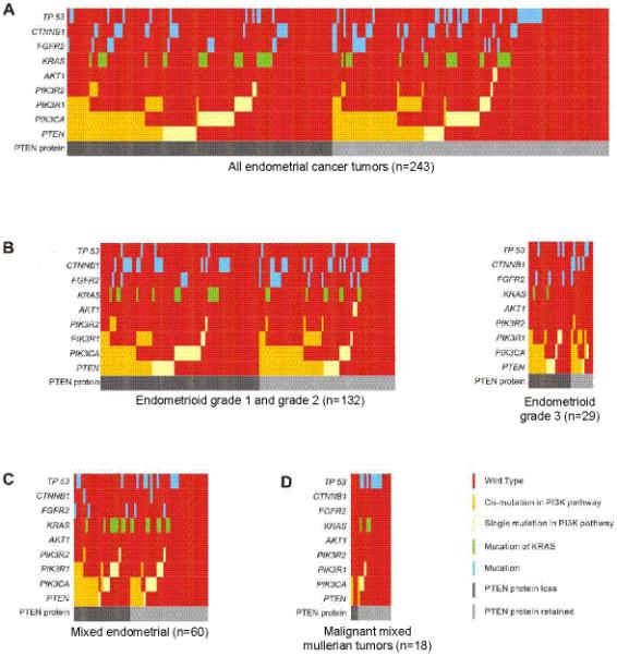
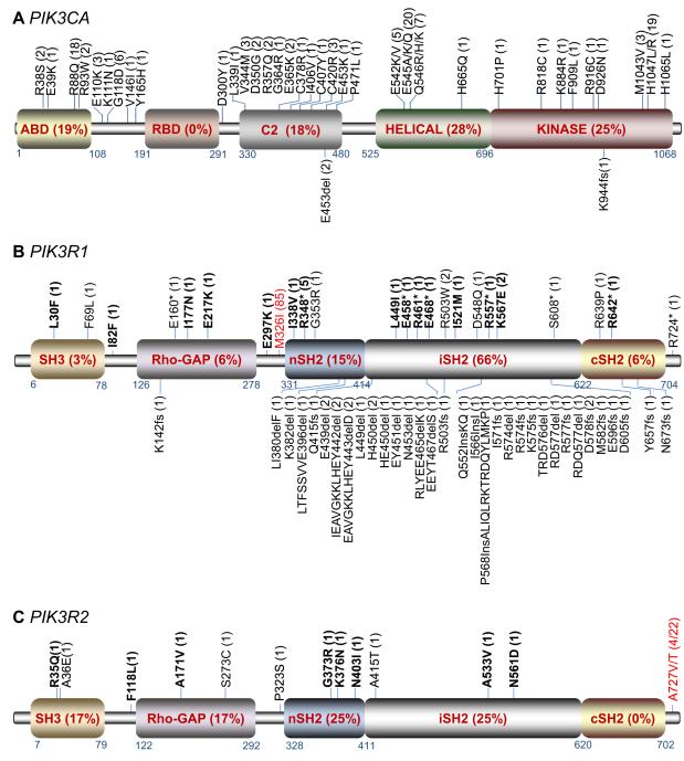
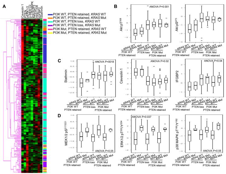
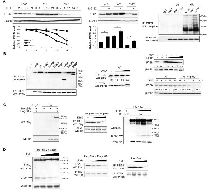
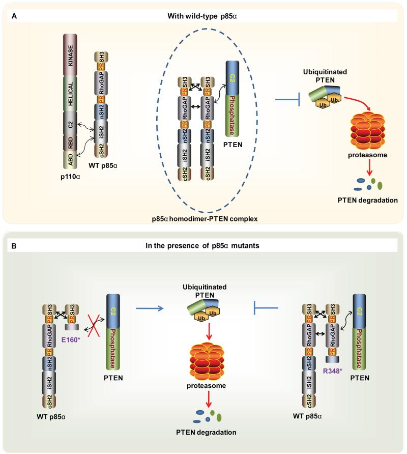
We demonstrate that phosphatidylinositol 3-kinase (PI3K) pathway aberrations occur in >80% of endometrioid endometrial cancers, with coordinate mutations of multiple PI3K pathway members being more common than predicted by chance. PIK3R1 (p85α) mutations occur at a higher rate in endometrial cancer than in any other tumor lineage, and PIK3R2 (p85β), not previously demonstrated to be a cancer gene, is also frequently mutated. The dominant activation event in the PI3K pathway appears to be PTEN protein loss. However, in tumors with retained PTEN protein, PI3K pathway mutations phenocopy PTEN loss, resulting in pathway activation. KRAS mutations are common in endometrioid tumors activating independent events from PI3K pathway aberrations. Multiple PIK3R1 and PIK3R2 mutations demonstrate gain of function, including disruption of a novel mechanism of pathway regulation wherein p85α dimers bind and stabilize PTEN. Taken together, the PI3K pathway represents a critical driver of endometrial cancer pathogenesis and a novel therapeutic target.

Keywords: Endometrial Cancer; PIK3CA; PIK3R1; PIK3R2; PTEN.


Figures

Figure 1
Figure 1
Mutation diagrams: A, the full set of endometrial tumor samples (n = 243); B, endometrioid grade 1 and grade 2 (n = 132; left), endometrioid grade 3 (n = 29; right); C, mixed endometrial (n = 60); and D, MMMT (n = 18). Each column represents a tumor and each row corresponds to a single gene.
Figure 2
Figure 2
Distribution of non-synonymous mutations in A, PIK3CA, B, PIK3R1 and C, PIK3R2. Substitutions (top) and indels (bottom) are graphed with the amino acid indicated and the number of independent observations within parentheses. The percentage of mutations identified in each domain is shown in the box. p110α is characterized by 5 domains: ABD, Ras-binding domain (RBD), C2 domain, helical domain, and kinase catalytic domain. p85 consists of SH3, Rho-GAP, and two SH2 domains (nSH2 and cSH2) that flank an intervening domain (iSH2). *, nonsense mutation; bold, mutations confirmed somatic; red, germline SNPs.
Figure 3
Figure 3
PI3K mutation in tumors where PTEN protein is retained mimics the effect of PTEN loss on downstream signaling regardless of KRAS status. A, heat map of unsupervised cluster analysis of proteins and samples by RPPA. Proteins are listed across the top of the heat map and mutational status of the tumor is represented at the right. Red, higher expression; green, low relative to the other samples. Expression levels of B, phosphorylated AKT at Thr308 (left) and Ser473 (right); C, stathmin (left), caveolin 1 (middle), insulin-like growth factor binding protein 2 (IGFBP2; right); D, phosphorylated MEK1/2 at Ser217/221 (left), ERK1/2 at Thr202/Tyr204 (middle), and p38 MAPK at Thr180/Tyr182 (right) were logarithmically converted, normalized by mean, and presented on the Y axis. The boxes represent the distribution of individual values from the lower 25th percentile to upper 75th percentile; solid line in the middle, median value; lower and upper whiskers, 5th and 95th percentiles. WT, wild-type; Mut, mutated.
Figure 4
Figure 4
Ba/F3 cells were transfected with A, wild-type p85α (p85α WT; left) or patient mutants, or p85β WT or patient mutants (right two gray bars, p85α WT or E160*). Cells were cultured without IL-3 for 4 weeks and harvested for viability assay. LacZ served as the control. The means (±SD) of triplicate samples of 3 independent experiments are shown. *, P < 0.05, compared with WT. The plasmids were transiently transfected into HEC1A cells. Whole-cell lysates were collected at 72 hours post-transfection for B, Western blot (WB) or C, immunoprecipitation (IP) with anti-p85α and then subjected to WB with anti-p110α. Numerical values below each lane of the immunoblots represent quantification of the relative protein level by densitometry (normalized to β-actin or AKT)
Figure 5
Figure 5
A, HEC1A cells transfected with LacZ or p85α wild-type (WT) or E160* were treated with cycloheximide (CHX; left) for indicated duration or MG132 (middle) for 24 hours. Cells were then harvested for Western blot (WB). PTEN levels were normalized to β-actin by densitometry (below). *, P < 0.05. Right, HEC1A cells were transfected with LacZ or WT or E160* in the absence or presence of ubiquitin (Ub) for 72 hours. Whole-cell lysates were collected for immunoprecipitation (IP) with anti-PTEN and then subjected to WB with anti-ubiquitin. PTEN protein levels were normalized prior to IP by using proportionally different amounts of lysates. B, left, HEC1A cells were transfected with LacZ or WT or its mutants for 72 hours and were collected for IP with PTEN and WB with anti-p85α. HEC1A cells were co-transfected with WT or increasing amount of E160*. Cell lysates were collected for IP (middle) or WB (right top). Right bottom, transfected HEC1A cells were treated with CHX for the indicated time points and harvested for WB. C, cells were transfected with HA-tagged p85α (HA-p85α) and/or Flag-tagged p85α (Flag-p85α) in the absence (left) or presence (middle) of increasing amount of E160*. IP was performed with anti-HA and WB with anti-Flag. Right, cells were transfected with HA-p85α and increasing amounts of E160*. IP was performed with anti-HA and WB with anti-p85α. D, left, cells were transfected with WT p110α, Flag-p85α and E160*. IP was performed with anti-Flag and WB with anti-p85α. Middle, cells were transfected with WT p110α, Flag-p85α and HA-p85α. IP was performed with anti-HA and WB with anti-Flag. Right, cells were transffected with WT p110α and HA-p85α. IP was performed with PTEN and WB with p85α. Numerical values below each lane of the immunoblots represent quantification of the relative protein level by densitometry.
Figure 6
Figure 6
Schematic working model of p85α-mediated PTEN stabilization. A, in cells with wild-type (WT) p85α, in addition to p110α-bound p85α, excess p85α forms homodimers via intermolecular interactions between SH3 and proline-rich motif and between Rho-GAP domains (see Discussion). The homodimer likely undergoes conformation changes that allow binding to PTEN and stabilizes PTEN through inhibition of ubiquitination. B, the E160* truncated mutant binds WT p85α but inhibits PTEN binding, resulting in PTEN degradation. However, the R348* mutant retains the ability to bind PTEN after dimerization, either alone or with WT p85α, thereby inhibiting PTEN degradation. PR, proline-rich motif.

Comment in

Similar articles

Cited by

References

    1. Hennessy BT, Smith DL, Ram PT, Lu Y, Mills GB. Exploiting the PI3K/AKT pathway for cancer drug discovery. Nat Rev Drug Discov. 2005;4:988–1004. - PubMed
    1. Daikoku T, Hirota Y, Tranguch S, Joshi AR, DeMayo FJ, Lydon JP, et al. Conditional loss of uterine Pten unfailingly and rapidly induces endometrial cancer in mice. Cancer Res. 2008;68:5619–27. - PMC - PubMed
    1. Mutter GL, Ince TA, Baak JP, Kust GA, Zhou XP, Eng C. Molecular identification of latent precancers in histologically normal endometrium. Cancer Res. 2001;61:4311–4. - PubMed
    1. Sun H, Enomoto T, Fujita M, Wada H, Yoshino K, Ozaki K, et al. Mutational analysis of the PTEN gene in endometrial carcinoma and hyperplasia. Am J Clin Pathol. 2001;115:32–8. - PubMed
    1. Kanamori Y, Kigawa J, Itamochi H, Shimada M, Takahashi M, Kamazawa S, et al. Correlation between loss of PTEN expression and Akt phosphorylation in endometrial carcinoma. Clin Cancer Res. 2001;7:892–5. - PubMed

Publication types

MeSH terms

Substances