. 2011 Dec;119(6):1294-305.
doi: 10.1111/j.1471-4159.2011.07517.x. Epub 2011 Nov 3.

Catecholamines up integrates dopamine synthesis and synaptic trafficking

Affiliations

Catecholamines up integrates dopamine synthesis and synaptic trafficking

Zhe Wang et al. J Neurochem. 2011 Dec.

Abstract

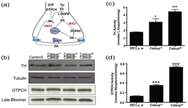
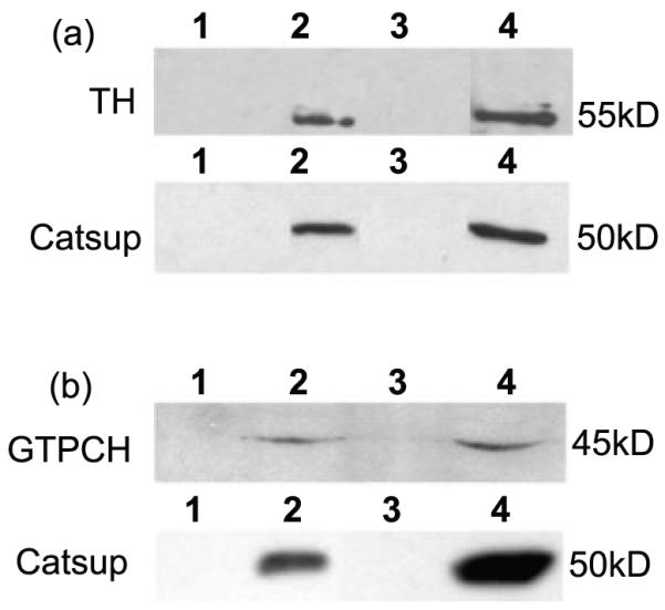
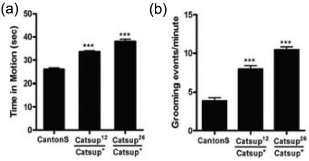
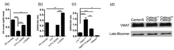
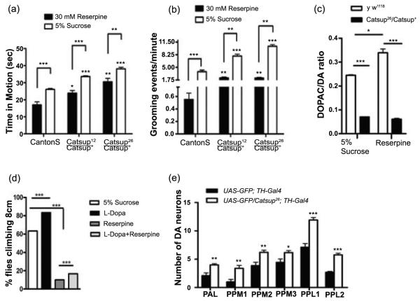
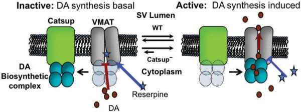
The highly reactive nature of dopamine renders dopaminergic neurons vulnerable to oxidative damage. We recently demonstrated that loss-of-function mutations in the Drosophila gene Catecholamines up (Catsup) elevate dopamine pools but, paradoxically, also confer resistance to paraquat, an herbicide that induces oxidative stress-mediated toxicity in dopaminergic neurons. We now report a novel association of the membrane protein, Catsup, with GTP cyclohydrolase rate-limiting enzyme for tetrahydrobiopterin (BH(4)) biosynthesis and tyrosine hydroxylase, rate-limiting enzyme for dopamine biosynthesis, which requires BH(4) as a cofactor. Loss-of-function Catsup mutations cause dominant hyperactivation of both enzymes. Elevated dopamine levels in Catsup mutants coincide with several distinct characteristics, including hypermobility, minimal basal levels of 3,4-dihydroxy-phenylacetic acid, an oxidative metabolite of dopamine, and resistance to the vesicular monoamine transporter inhibitor, reserpine, suggesting that excess dopamine is synaptically active and that Catsup functions in the regulation of synaptic vesicle loading and release of dopamine. We conclude that Catsup regulates and links the dopamine synthesis and transport networks.


Figures

Figure 1
Figure 1
Catsup mutations dominantly elevate TH and GTPCH activity through a post-translational mechanism. (a) Key components of DA synthesis, transport and metabolism. GTP is converted via three enzymatic reactions to the cofactor BH4. GTPCH catalyzes the rate-limiting step. TH converts tyrosine (tyr) to L-DOPA, in a BH4-dependent reaction. Aromatic amino acid decarboxylase subsequently converts L-DOPA to DA. DA is then transported into synaptic vesicles via VMAT or is converted by a two-step pathway into DOPAC by Monoamine Oxidase (MAO) and aldehyde dehydrogenase. After synaptic release, residual extracellular DA may be transported by into the pre-synaptic terminus by Dopamine Transporter (DAT) or may interact with the D2 autoreceptor (D2R) to trigger a G-protein coupled signaling pathway that leads to dephosphorylation of TH, which down-regulates its activity. (b) Immunoblot analysis of 3-5 day old flies was performed using antibodies against dTH, dGTPCH and loading controls, ß-tubulin and late-bloomer. Levels of TH and GTPCH protein are indistinguishable in the wild type (Canton S) and heterozygous Catsup mutant head extracts. (c, d) Extracts of adult male and female heads (in equal numbers), collected 24-48 hrs post-eclosion, were assayed for TH and GTPCH activity. Activities are expressed as nmoles of product per min. per mg protein. TH and GTPCH activities are elevated in head extracts of Catsup26/ Catsup+ and Catsup12/Catsup+ mutants relative to the wild type control (*, p< 0.05, **, p<0.01, *** p<0.001, n=5).
Figure 2
Figure 2
Catsup co-localizes in cell bodies and synaptic regions of dopaminergic neurons with TH and VMAT. (a-g) Co-localization (yellow, b) of TH (green, a) and Catsup (red, c) in adult CNS, detected with anti-dTH and anti-Catsup antibodies. (a-c) TH and Catsup co-expressed in DA neuron clusters in the central brain. Scale bar = 100 μm. (d,g) enlargements of indicated regions of (c) showing co-expression in cell bodies and synaptic termini. Scale bars = 50 μm. (e and f) Enlargements of (d) and (g), respectively, showing further details of synaptic co-localization of TH and Catsup. Scale bars = 10 μm. (h-l) Co-localization (yellow, i) of VMAT (green, h) and FLAG::Catsup (red, j) in CNS of w; TH-Gal4; UAS-FLAG::Catsup adults, detected with anti-VMAT and anti-FLAG antibodies. (h-j) VMAT and FLAG::Catsup expression in the central brain, Catsup is densely expressed in the lower brain as well as in specific regions in the central brain. Scale bars = 50 μm (k, l) Enlargements of indicated regions of (i), showing Catsup and VMAT co-localization within intracellular compartments (k, arrow heads) and in synaptic bouton-like puncta (l, arrowheads). Scale bars 20 μm.
Figure 3
Figure 3
TH and GTPCH co-immunoprecipitate with Catsup. (a) TH interacts with Catsup. (b) GTPCH interacts with Catsup. The upper panels in (a) and (b) show the results of anti-Catsup immunoprecipitation from head extracts; the blots were probed with anti-dTH (a) and anti-GTPCH (b). The lower panels shows the results of anti-dTH (a) and anti-GTPCH (b) immunoprecipation from head extracts; the blots were probed with anti-Catsup antibody. The lanes in each panel are in the following order: Lane 1- Beads with no antibody + extract- a control for non-specific retention of proteins. Lane 2- Co-immunoprecipitated protein Lane 3: blank. 4: Extract only-detecting endogenous proteins of the same molecular mass as the co-precipitated proteins in lanes 2.
Figure 4
Figure 4
Amine-linked mobility is elevated in heterozygous Catsup mutants. (a) Overall activity measured as the portion of 45 sec intervals that each of 20 adult males of each genotype (3-5 days post-eclosion) was in motion. Both Catsup12/Catsup+ and Catsup26/Catsup+, were hyperactive relative to wild type (Canton S) flies. (b) Grooming, quantified as the number of grooming events in 60 sec intervals, was determined using 20 adult male flies (3-5 days post-eclosion). Catsup mutants had higher grooming frequencies than the wild type flies. Statistical analysis was performed using one-way ANOVA. Error bars represent mean SEM. (*** p< 0.001).
Figure 5
Figure 5
Pharmacological and genetic manipulation of cytosolic dopamine pools. (a, b) Pharmacological modulation of DA levels in y w1118 flies. Adults, 48 h post-eclosion, were fed 5% sucrose, 30 mM 3-iodo-tyrosine (3-IT), 30 mM 3-IT + 1mM L-DOPA, or 1mM L-DOPA for 12 hrs. (a) DA levels were significantly depleted in the presence of 3-IT, and significantly elevated when fed L-DOPA. Co-feeding 3-IT and L-DOPA resulted in near normal DA pools. (b) DOPAC:DA ratios respond to pharmacological alteration of DA pools. The TH inhibitor, 3-IT, reduced the DOPAC:DA ratio to a negligible level, while flies treated with L-DOPA had significantly higher DOPAC:DA ratios relative to controls. These results indicate that removal of DA from the cytosol is not solely dependent on cytosolic DA levels. (c) Double mutant analysis of DOPAC:DA ratios in VMATD14/Catsup26 reveals a functional interaction. VMATD1 /VMAT+ and Catsup26/Catsup+ mutants display strongly elevated and reduced DOPAC:DA ratios, respectively, in comparison to the y w1118 flies. The transheterozygous combination of Catsup26 and VMATD14 mutations partially rescues the DOPAC:DA ratio relative to VMATD1 /VMAT+ flies. Statistical analysis was performed using one-way ANOVA. Error bars represent mean SEM. (*, p<0.05, **, p<0.01, ***, p<0.001, n=5). (d) Mutations in Catsup do not alter VMAT protein levels. Immunoblot analysis was performed using antibodies against dVMAT and loading control, Late-bloomer.
Figure 6
Figure 6
Administration of VMAT inhibitor, reserpine, reveals a functional interaction between VMAT and Catsup. Mutations in Catsup diminish the effects of reserpine on overall mobility (a) and grooming (b) relative to wild type controls. Reserpine reduced mobility in all cases, but was unable to restore mobility to a wild type level in Catsup mutants. (c) Reserpine ingestion causes an increase in the DOPAC:DA ratio of wild type, but not Catsup26/Catsup+ mutant, heads. (a-c) Adult males of each genotype were fed 30 mM reserpine in 5% sucrose. N = 20 for each test and genotype. (d) Elevation of DA pools alone (in absence of Catsup mutation) does not rescue mobility from the effects of reserpine. Adult males were fed 1 mM L-DOPA and/or 10 mM reserpine for 12 hrs, after a 12 hr pre-feeding of 1 mM L-DOPA. Mobility was assayed as the percent of the flies assayed in three replicas that were able to climb 8 cm in 4 sec. Each replica consisted of 5 adult males per vial, 10 vials. (e) Acute exposure to reserpine (120 mM, 48 hrs) causes dopaminergic neurodegeneration in Catsup+, but not Catsup26/Catsup+ flies. Both Catsup wild type and mutants were combined with UAS-GFP/+; TH-Gal4/+ to visualize dopaminergic neurons. Individual dopaminergic clusters were scored in 10-12 brains per genotype. PAL (protocerebral anterolateral), PPM1, PPM2, PPM3 (protocerebral posterior medial), PPL1 and PPL2 (protocerebral posterolateral) neurons were counted individually. Wild type brains displayed significantly greater loss of DA neurons in all clusters, relative to Catsup mutant brains. The significance of differences between Catsup mutant and wild type neurons in each cluster *** p<0.0001; ** p <0.001 and * p<0.05.
Figure 7
Figure 7
Model for functional interaction between Catsup and VMAT. Catsup is located either in the synaptic vesicle membrane or in another membrane in close proximity. The BH4 and DA biosynthetic complexes are proposed to interact with Catsup in the absence of neuron stimulation. In this state, N-terminal regulatory domains of TH and GTPCH are blocked from phosphorylation by ser/thr kinases, minimizing biosynthetic activity. Upon neuron stimulation, Catsup releases the complex, which then may undergo phosphorylation and interaction with VMAT, allowing efficient vesicular transport of DA. In the inactive state, reserpine has ready access to its binding site on VMAT, preventing DA transport. In the active state, reserpine access is partially blocked. In Catsup mutants, diminished levels of Catsup result in increased interactions between the biosynthesis complex and VMAT, prolonging activation and blockade of reserpine binding.

References

    1. Asanuma M, Miyazaki I, Ogawa N. Dopamine- or L-DOPA-induced neurotoxicity: The role of dopamine quinone formation and tyrosinase in a model of Parkinson’s disease. Neurotox Res. 2003;5:165–76. - PubMed
    1. Bayersdorfer F, Voigt A, Schneuwly S, Botella JA. Dopamine-dependent neurodegeneration in Drosophila models of familial and sporadic Parkinson’s disease. Neurobiol Dis. 2010 In Press. - PubMed
    1. Berman SB, Hastings TG. Dopamine oxidation alters mitochondrial respiration and Induces permeability transition in brain mitochondria: Implications for Parkinson’s disease. J Neurochem. 1999;73:1127–1137. - PubMed
    1. Bonini NM, Fortini ME. Human neurodegenerative disease modeling using Drosophila. Annu. Rev. Neurosci. 2003;26:627–656. - PubMed
    1. Bowling KM, Huang Z, Xu D, Funderburk CD, Karnik N, Ferdousy F, Neckameyer W, O’Donnell JM. Direct binding of GTP cyclohydrolase and tyrosine hydroxylase: Regulatory interactions between key enzymes in dopamine biosynthesis. J Biol Chem. 2008;283:31449–31459. - PMC - PubMed

Publication types

MeSH terms