. 2011 Nov;166(2):258-68.
doi: 10.1111/j.1365-2249.2011.04458.x.

Suppressive role of hepatic dendritic cells in concanavalin A-induced hepatitis

Affiliations

Suppressive role of hepatic dendritic cells in concanavalin A-induced hepatitis

C Tomiyama et al. Clin Exp Immunol. 2011 Nov.

Abstract

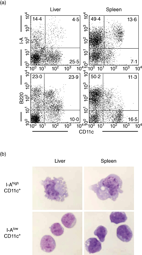
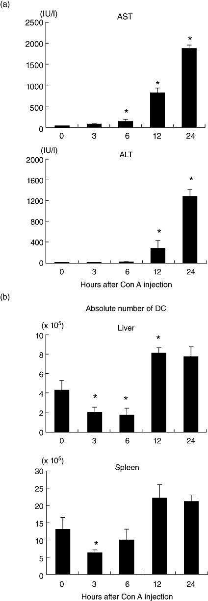
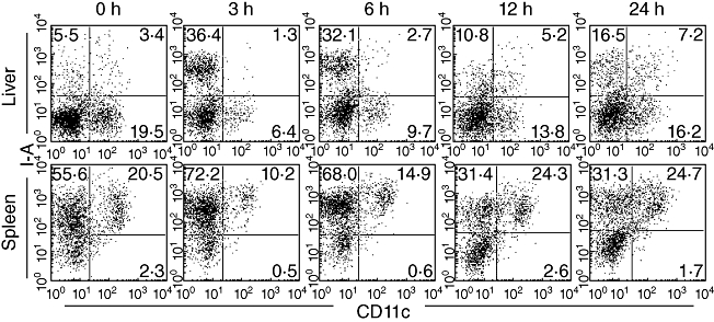
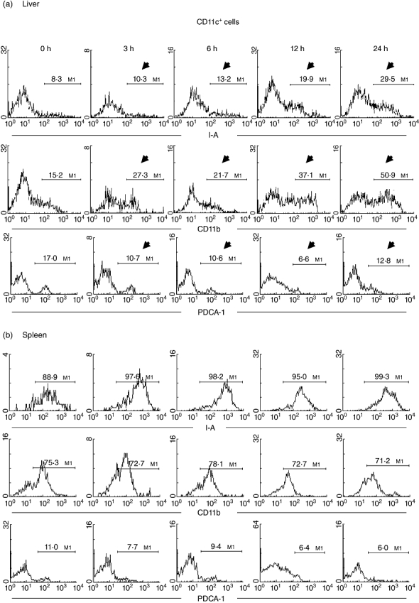
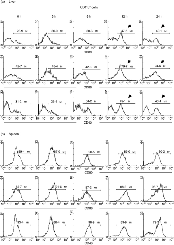
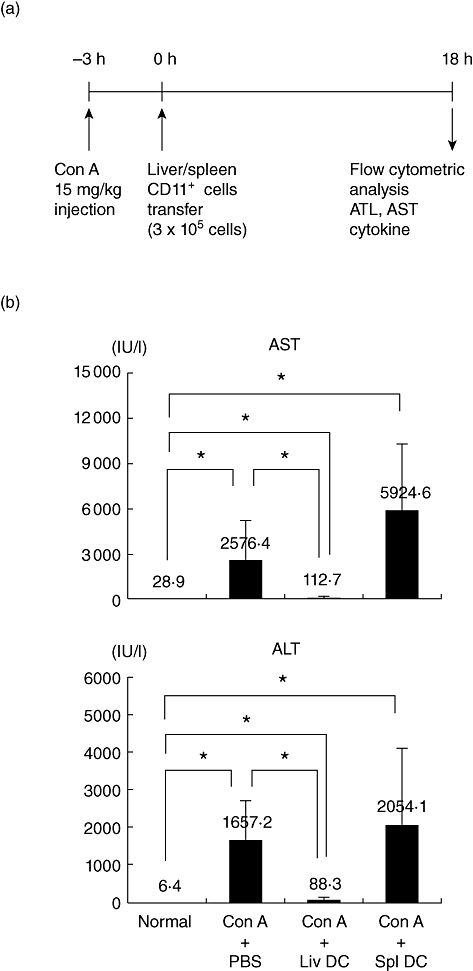
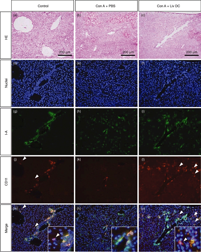
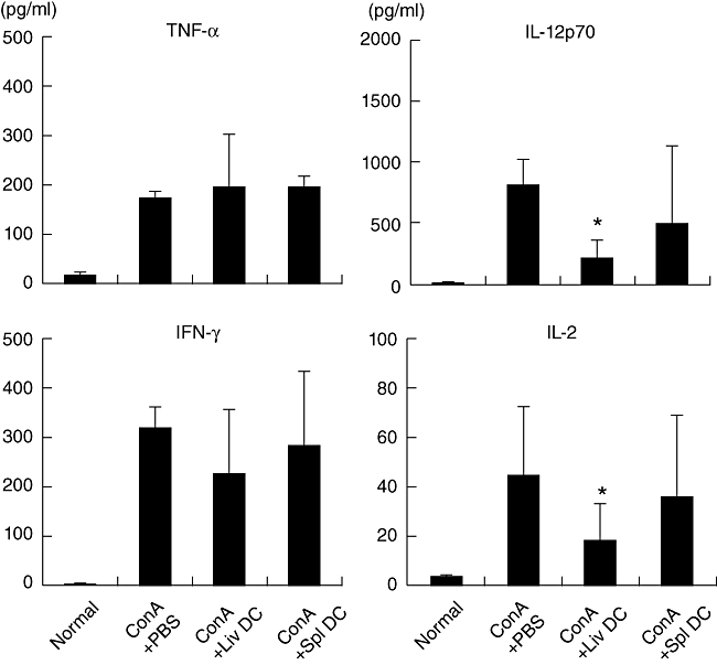
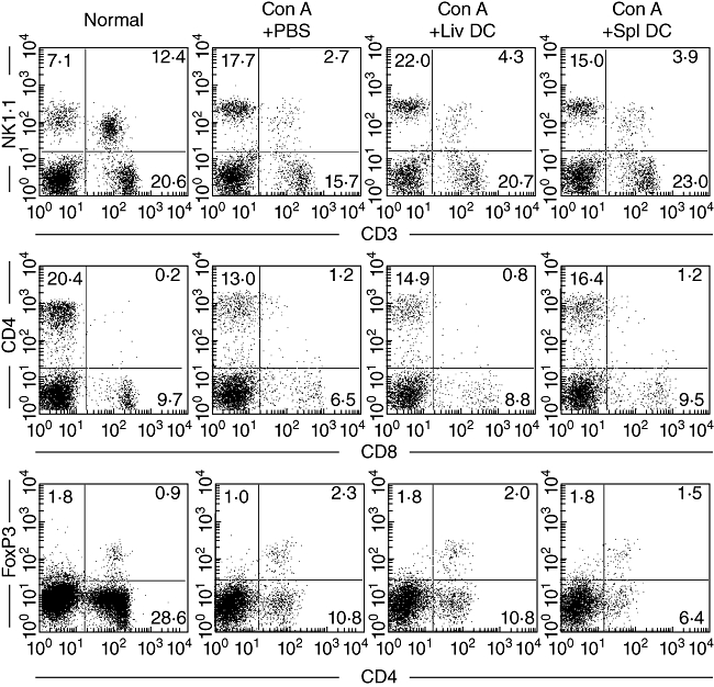
Concanavalin A (Con A)-induced hepatitis is a mouse model of acute autoimmune hepatitis. The aim of this study was to investigate the role of hepatic dendritic cells (DC) in the immune modulation of tissue damage. Almost all hepatic DC were plasmacytoid DC (CD11c+ I-A(low) B220+); however, conventional DC were CD11c+ I-A(high) B220(-). At an early stage (3-6 h) after Con A administration, the number of DC in both the liver and spleen decreased, increasing thereafter (12-24 h) in parallel with hepatic failure. The hepatic CD11c+ DC population contained many CD11b(-) cells, while the majority of splenic CD11c+ DC were CD11b+. After Con A administration, the proportion of I-A+ and CD11b+ cells within the CD11c+ DC population tended to increase in the liver, but not in the spleen. Similarly, expression of the activation markers CD80, CD86 and CD40 by CD11c+ DC increased in the liver, but not in the spleen. Next, adoptive transfer of DC isolated from the liver and spleen was performed 3 h after Con A administration to examine the immunomodulatory function of DC. Only hepatic DC had the ability to suppress hepatic failure. Analysis of cytokine production and subsequent identification of the effector cells showed that hepatic DC achieved this by suppressing the production of interleukin (IL)-12 and IL-2, rather than modulating effector cell function.


Figures

Fig. 1
Fig. 1
Characterization of dendritic cells (DC) in the liver and spleen. (a) Phenotype and (b) morphology. DC phenotype was characterized by immunofluorescence analysis. The expression of I-A and B220 on CD11c+ DC was examined. I-Ahigh CD11c+ and I-Alow CD11c+cells were purified using a cell sorter and then stained using the Giemsa method. Representative data of four experiments are depicted.
Fig. 2
Fig. 2
Kinetics of tissue damage and the number of dendritic cells (DC) after concanavalin A administration. (a) The levels of aspartate aminotransferase (AST) and alanine aminotransferase (ALT) in sera. (b) The absolute numbers of CD11c+ DC cells in the liver and spleen. The mean ± 1 standard error were produced by five experiments.
Fig. 3
Fig. 3
Variations in the proportion of I-A+ CD11c+ dendritic cells (DC) in the liver and spleen after concanavalin A administration. Two-colour staining for CD11c and I-A was conducted. Data are representative of three independent experiments.
Fig. 4
Fig. 4
Further characterization of dendritic cell (DC) phenotype in (a) liver and (b) spleen. Two-colour staining for CD11c and I-A (or CD11b or PDCA-1) was conducted. Gated analysis was used to examine the proportion of DC expressing I-A, CD11b or PDCA-1. Significant changes are indicated by arrows. Representative data of five experiments are depicted.
Fig. 5
Fig. 5
Further characterization of dendritic cell (DC) phenotype in (a) liver and (b) spleen. Two-colour staining for CD11c and CD80 (or CD86 or CD40) was conducted. Gated analysis was used to examine the proportion of DC expressing CD80, CD86 and CD40. Representative data of five experiments are depicted. Significant changes are indicated by arrows.
Fig. 6
Fig. 6
Adoptive transfer of dendritic cells (DC). (a) Time schedule and (b) modulation of tissue damage by adoptive transfer of DC. DC were transferred 3 h after concanavalin A administration. Mean ± 1 standard error were produced by three experiments. *P < 0·05.
Fig. 7
Fig. 7
Histology and immunohistochemical staining of the liver. (a, d, g, j, m) Control mice. (b, e, h, k, n) Mice treated with concanavalin A (Con A). (c, f, i, l, o) Mice treated with Con A plus adoptive transfer of liver dendritic cells (DC). (a–c) Haematoxylin and eosin staining. (d–o) Immunohistochemical staining. White arrowheads indicate the CD11c single positive cells. They were only seen in the control liver and the DC-post injected Con A-treated liver, but not in the Con A-treated liver. Representative data of three experiments are depicted.
Fig. 8
Fig. 8
Cytokine production after adoptive transfer of dendritic cells (DC). The level of cytokines was determined 24 h after concanavalin A administration. Data represent the mean ± 1 standard error from four experiments. *P < 0·05.
Fig. 9
Fig. 9
Identification of lymphocyte subsets by immunofluorescence analysis. Natural killer (NK) T cells were NK1·1+ CD3int. CD4+ T cells were CD4+ CD8. Regulatory T cells were CD4+ forkhead box P3+. Data are representative of three independent experiments.

References

    1. Jomantaite I, Dikopoulos N, Kröger A, et al. Hepatic dendritic cell subsets in the mouse. Eur J Immunol. 2004;34:355–65. - PubMed
    1. Lau AH, Thomson AW. Dendritic cells and immune regulation in the liver. Gut. 2003;52:307–14. - PMC - PubMed
    1. Kingham TP, Chaudhry UI, Plitas G, Katz SC, Raab J, DeMatteo RP. Murine liver plasmacytoid dendritic cells become potent immunostimulatory cells after Flt-3 ligand expansion. Hepatology. 2007;45:445–54. - PubMed
    1. Pillarisetty VG, Shah AB, Miller G, Bleier JI, DeMatteo RP. Liver dendritic cells are less immunogenic than spleen dendritic cells because of differences in subtype composition. J Immunol. 2004;172:1009–17. - PubMed
    1. Castellaneta A, Sumpter TL, Chen L, Tokita D, Thomson AW. NOD2 ligation subverts IFN-α production by liver plasmacytoid dendritic cells and inhibits their T cell allostimulatory activity via B7-H1 up-regulation. J Immunol. 2009;183:6922–32. - PMC - PubMed

Publication types

MeSH terms