. 2011 Nov;166(2):281-90.
doi: 10.1111/j.1365-2249.2011.04471.x.

Interleukin-17 exacerbates hepatic steatosis and inflammation in non-alcoholic fatty liver disease

Affiliations

Interleukin-17 exacerbates hepatic steatosis and inflammation in non-alcoholic fatty liver disease

Y Tang et al. Clin Exp Immunol. 2011 Nov.

Abstract

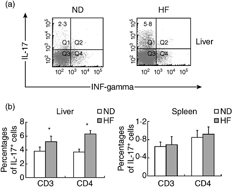
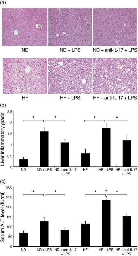
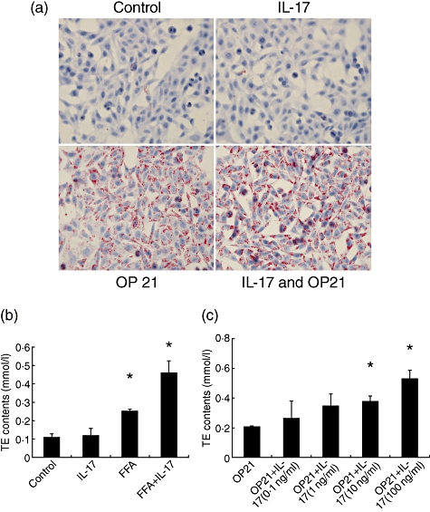
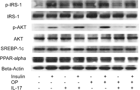
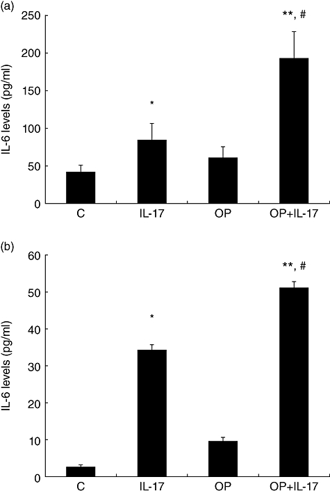
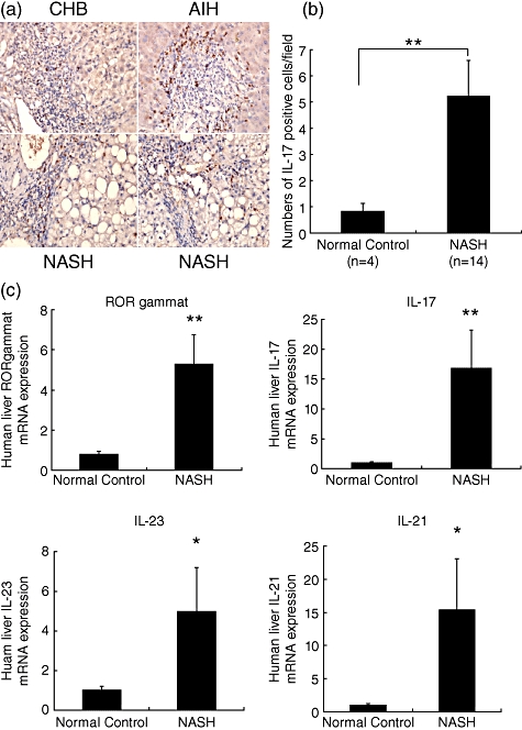
Mechanisms associated with the progression of simple steatosis to non-alcoholic fatty liver disease (NAFLD) remain undefined. Regulatory T cells (T(regs)) play a critical role in regulating inflammatory processes in non-alcoholic steatohepatitis (NASH) and because T helper type 17 (Th17) functionally oppose T(reg)-mediated responses, this study focused on characterizing the role of Th17 cells using a NAFLD mouse model. C57BL/6 mice were fed either a normal diet (ND) or high fat (HF) diet for 8 weeks. Mice in the HF group had a significantly higher frequency of liver Th17 cells compared to ND-fed mice. Neutralization of interleukin (IL)-17 in HF mice ameliorated lipopolysaccharide (LPS)-induced liver injury reflected by decreased serum alanine aminotransferase (ALT) levels and reduced inflammatory cell infiltrates in the liver. In vitro, HepG2 cells cultured in the presence of free fatty acids (FFA; oleic acid and palmitic acid) for 24 h and IL-17 developed steatosis via insulin-signalling pathway interference. IL-17 and FFAs synergized to induce IL-6 production by HepG2 cells and murine primary hepatocytes which, in combination with transforming growth factor (TGF-β), expanded Th17 cells. It is likely that a similar process occurs in NASH patients, as there were significant levels of IL-17(+) cell infiltrates in NASH patient livers. The hepatic expression of Th17 cell-related genes [retinoid-related orphan receptor gamma (ROR)γt, IL-17, IL-21 and IL-23] was also increased significantly in NASH patients compared to healthy controls. Th17 cells and IL-17 were associated with hepatic steatosis and proinflammatory response in NAFLD and facilitated the transition from simple steatosis to steatohepatitis. Strategies designed to alter the balance between Th17 cells and T(regs) should be explored as a means of preventing progression to NASH and advanced liver diseases in NAFLD patients.


Figures

Fig. 1
Fig. 1
Increase in T helper type 17 (Th17) cells in the livers of mice fed high fat (HF) diets. C57BL/6 mice were fed normal diet (ND) or HF diets for 8 weeks. Hepatic mononuclear cells (HMNCs) were isolated and incubated with labelled antibodies specific to CD3 or CD4 as well as antibodies specific for intracellular interleukin (IL)-17 or interferon (IFN)-γ. Total liver mononuclear cell numbers were similar for mice fed either diet. (a) Representative dot-plots of gated CD4+ cells. (b) Mean [standard deviation (s.d.)] values of intracellular IL-17+ IFN-γ- staining HMNC (gated CD3+ or CD4+ cells) from murine livers or spleens (n = 6 in each group, *P < 0·05).
Fig. 2
Fig. 2
Neutralization of interleukin (IL)-17 attenuates lipopolysaccharide (LPS)-induced liver injury in high fat (HF) diet mice. C57BL/6 mice were fed a normal diet (ND) or HF diet for 8 weeks. Mice were injected intraperitoneally with a single dose of LPS (50 µg/mouse). Neutralizing IL-17 antibody (100 µg/mice) was administrated intravenously in some mice from each group prior to LPS injection. Serum and liver tissue samples were obtained 6 h after LPS injection. (a) Representative haematoxylin and eosin-stained liver sections (magnification ×200) from ND and HF mice. (b) Mean [standard deviation (s.d.)] liver inflammatory grade of triplicate experiments (n = 6/experiment). (c) Mean (s.d.) serum alanine aminotransferase (ALT) levels of triplicate experiments (n = 6/experiment). *P ≤ 0·05, #P ≤ 0·05 versus ND mice treated with LPS.
Fig. 3
Fig. 3
Neutralization of IL-17 attenuates lipopolysaccharide (LPS)-induced mitogen-activated protein kinase (MAPK) and nuclear factor (NF)-κB signalling pathways in normal diet (ND) and high fat (HF) mice. Animal experiments were performed as described in Fig. 2. Western blots were performed on whole liver protein extracts with antibodies specific to phosphorylated c-Jun N-terminal kinase (JNK) and on nuclear extracts to detect activation of the NF-κB signalling pathway [inhibitor of nuclear factor kappa-B (IKBα)]. Membranes were then stripped and reprobed with antibodies specific to JNK and IKBα. β-actin expression was used a loading control. (a) Representative autoradiographs of JNK and IKBα. (b) Representative autoradiographs of NF-κB binding activity from liver nuclear extracts.
Fig. 4
Fig. 4
Interleukin (IL)-17 exacerbated hepatocyte steatosis in HepG2 cells in vitro. The free fatty acids (FFA) mixture (oleate acid and palmitate acid at 1:2 ratio, OP21) was added to HepG2 cells to induce fat overloading for 24 h. IL-17 (100 ng/ml) was added to the culture medium with or without FFA for 24 h. (a) Representative Oil-Red staining of HepG2 cells treated with FFA with or without IL-17. (b) Intracellular triglyceride (TG) content was determined using an enzymatic kit. Mean [standard deviation (s.d.)] intracellular TG content of triplicate experiments (n = 4/experiment). *P < 0·05 versus controls. (c) The OP21 was added to HepG2 cells with different dosages of IL-17 (0·1–100 ng/ml) to induce fat overloading for 24 h. Intracellular TG content was determined using an enzymatic kit. *P < 0·05 versus OP21 treated without IL-17 group.
Fig. 5
Fig. 5
Interleukin (IL)-17 interfered with the insulin-signalling pathway in vitro. Representative autoradiographs of insulin pathway [insulin substrate receptor 1 (IRS-1) and protein kinase B (AKT) and fatty acid metabolism [sterol receptor element-binding protein-1c and peroxisome proliferator-activated receptor-α (SREBP-1c and PPAR-α)].
Fig. 6
Fig. 6
Interleukin (IL)-6 production by HepG2 cells and murine primary hepatocytes. IL-6 levels in HepG2 supernatants were measured by enzyme-linked immunosorbent assay (ELISA). (a) Mean [standard deviation (s.d.)] IL-6 levels in the supernatants of HepG2 cells treated with IL-17 or/and free fatty acid (FFA) (n = 4/experiment). (b) Mean [standard deviation (s.d.)] IL-6 levels in the supernatants of murine primary hepatocytes treated with IL-17 or/and FFA (n = 4/experiment), *P < 0·05 versus control; **P < 0·01 versus control; #P < 0·05 versus IL-17 group.
Fig. 7
Fig. 7
Interleukin (IL)-17 and other T helper type 17 (Th17)-related cytokine profile analysis of human liver tissues. (a) Representative liver staining for the presence of Th17 cells (IL-17+, brown-stained cells, magnification (×400). (b) Mean [standard deviation (s.d.)] numbers of IL-17 positive cells/field. Liver tissues were obtained from healthy controls (HC, n = 4) or from non-alcoholic steatohepatitis (NASH, n = 14) patients. Th17 cells in the liver were evaluated by immunohistochemical staining of IL-17. (c) Mean (s.d.) expression levels of the Th17-related cytokines retinoid-related orphan receptor (ROR)γt, IL-17, IL-21 and IL-23. Th17-related cytokines expressions were determined by quantitative reverse transcription–polymerase chain reaction (qRT–PCR) and normalized against glyceraldehyde 3-phosphate dehydrogenase (GAPDH) expression. Liver tissues were obtained from healthy controls (HC, n = 5) or NASH (n = 8) patients. *P < 0·05; **P < 0·01 compared with normal controls.

References

    1. Farrell GC, Larter CZ. Nonalcoholic fatty liver disease: from steatosis to cirrhosis. Hepatology. 2006;43(Suppl. 1):S99–S112. - PubMed
    1. Kleiner DE, Brunt EM, Van Natta M, et al. Design and validation of a histological scoring system for nonalcoholic fatty liver disease. Hepatology. 2005;41:1313–21. - PubMed
    1. Brunt EM. Nonalcoholic steatohepatitis. Semin Liver Dis. 2004;24:3–20. - PubMed
    1. Cortez-Pinto H, de Moura MC, Day CP. Non-alcoholic steatohepatitis: from cell biology to clinical practice. J Hepatol. 2006;44:197–208. - PubMed
    1. Kremer M, Hines IN, Milton RJ, Wheeler MD. Favored T helper 1 response in a mouse model of hepatosteatosis is associated with enhanced T cell-mediated hepatitis. Hepatology. 2006;44:216–27. - PubMed

Publication types

MeSH terms