. 2011 Oct 10:10:127.
doi: 10.1186/1476-4598-10-127.

L1CAM protein expression is associated with poor prognosis in non-small cell lung cancer

Affiliations

L1CAM protein expression is associated with poor prognosis in non-small cell lung cancer

Verena Tischler et al. Mol Cancer. .

Abstract

Background: The L1 cell adhesion molecule (L1CAM) is potentially involved in epithelial-mesenchymal transition (EMT). EMT marker expression is of prognostic significance in non-small cell lung cancer (NSCLC). The relevance of L1CAM for NSCLC is unclear. We investigated the protein expression of L1CAM in a cohort of NSCLC patients. L1CAM protein expression was correlated with clinico-pathological parameters including survival and markers of epithelial-mesenchymal transition.

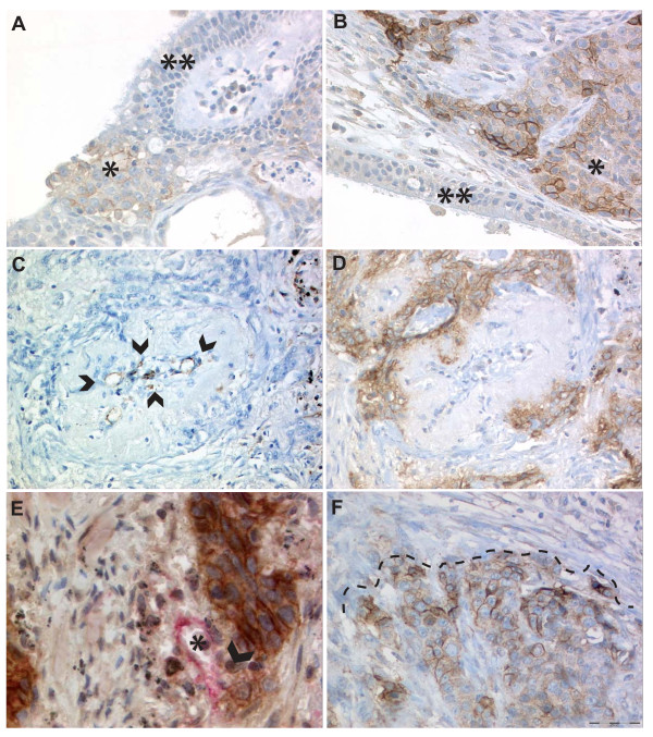
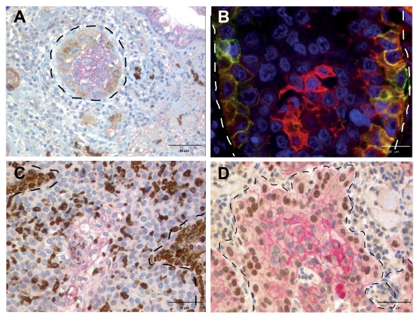
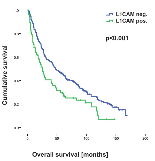
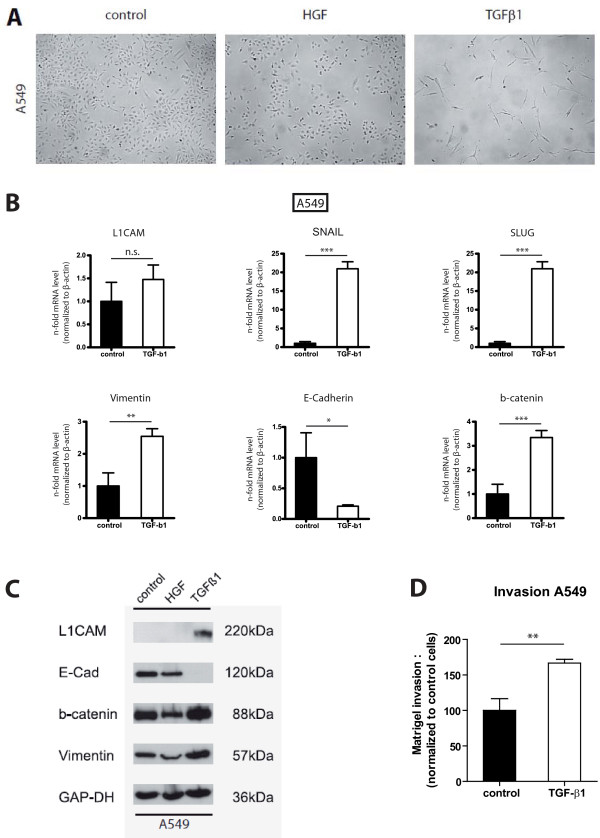
Results: L1CAM protein expression was found in 25% of squamous cell carcinomas and 24% of adenocarcinomas and correlated with blood vessel invasion and metastasis (p < 0.05). L1CAM was an independent predictor of survival in a multivariate analysis including pT, pN, and pM category, and tumor differentiation grade. L1CAM expression positively correlated with vimentin, beta-catenin, and slug, but inversely with E-cadherin (all p-values < 0.05). E-cadherin expression was higher in the tumor center than in the tumor periphery, whereas L1CAM and vimentin were expressed at the tumor-stroma interface. In L1CAM-negative A549 cells the L1CAM expression was upregulated and matrigel invasion was increased after stimulation with TGF-beta1. In L1CAM-positive SK-LU-1 and SK-LC-LL cells matrigel invasion was decreased after L1CAM siRNA knockdown.

Conclusions: A subset of NSCLCs with vessel tropism and increased metastasis aberrantly expresses L1CAM. L1CAM is a novel prognostic marker for NSCLCs that is upregulated by EMT induction and appears to be instrumental for enhanced cell invasion.


Figures

Figure 1
Figure 1
Expression of L1CAM in NSCLC. A/B) SCC with expression of L1CAM (*) whereas the bronchial ciliated and focally metaplastic epithelium is negative (**), magnification 200x. Note on Figure 1B the pronounced expression of L1CAM in some areas of the tumor-stroma interface. C/D) Serial section, C CD31 (endothelial cells marked by arrowheads) and D L1CAM, of a SCC surrounding and partially invading the media of a blood vessel. Note that intratumoral vessels regularly show profound remodeling of the arteriolar wall with disappearance of the elastic layers normally bordering the media myocytes, magnification 200x. E) L1CAM positive tumor cells (brown) destroying the vessel wall lined by CD31 positive (red) endothelial cells. F) Accentuated L1CAM expression at the tumor-stroma interface (dashed line) and weaker (heterogeneous) L1CAM expression in the lower part of the picture representing a central part of the tumor. Hematoxylin counterstain was used for all slides.
Figure 2
Figure 2
L1CAM and EMT marker expression patterns at the tumor-stroma interface. A) SCC with expression of L1CAM at the tumor-stroma interface (brown). The tumor center shows moderate E-cadherin expression (red). At the tumor-stroma interface, E-cadherin expression is decreased. Note the strong positivity of small peripheral nerves for L1CAM. B) Double IF staining for L1CAM (green) and E-cadherin (red): L1CAM is expressed at the tumor border and E-cadherin expression is strongest in the tumor center. E-cadherin expression is decreased at the tumor border (yellow). C) Membranous E-cadherin (red) is expressed in the tumor center and decreased towards the tumor-stroma interface. Two strong Vimentin positive (brown) stromal cell aggregates are marked with dotted lines (upper left and mid to lower right). Note that most of the Vimentin positive cells show nuclear morphology of the tumor cells. CD68 staining to exclude Vimentin positive macrophages was not performed. D) Decrease of membranous E-cadherin (red) and strong nuclear expression of slug (brown) at the tumor-stroma interface of a SCC. In the tumor center E-cadherin is strongly but slug is not expressed (blue nuclei). Hematoxylin and DAPI counterstain were used, respectively. Tumor-stroma interfaces are marked by dotted lines.
Figure 3
Figure 3
L1CAM expression is correlated with shortened overall survival.
Figure 4
Figure 4
L1CAM in A549. A) Stimulation of A549 cells with TGF-beta1 induces a mesenchymal phenotype whereas HGF does not alter cell morphology. Note that TGF-beta1 treatment affected the proliferation of A549 cells. B/C) A549 cells stimulated with TGF-beta1 show an EMT phenotype and increased L1CAM expression on mRNA and protein level. One representative experiment of n = 3 is shown. GAP-DH loading control. D) Matrigel invasion of A549 cells is enhanced by TGF-beta1 stimulation. The error bars represent mean values ± SEM. One representative experiment of n = 3 is shown.
Figure 5
Figure 5
SiRNA knockdown reduces matrigel invasion. A) SiRNA knockdown suppresses L1CAM expression in SK-LU-1 and SK-LC-LL cells. B) L1CAM siRNA knockdown significantly reduces matrigel invasion of SK-LU-1 and SK-LC-LL cells. The error bars represent mean values ± SEM. One representative experiment of n = 3 is shown.

References

    1. Lindner J, Rathjen FG, Schachner M. L1 mono- and polyclonal antibodies modify cell migration in early postnatal mouse cerebellum. Nature. 1983;305:427–430. doi: 10.1038/305427a0. - DOI - PubMed
    1. Schafer MK, Altevogt P. L1CAM malfunction in the nervous system and human carcinomas. Cell Mol Life Sci. 2010;67:2425–2437. doi: 10.1007/s00018-010-0339-1. - DOI - PMC - PubMed
    1. Moos M, Tacke R, Scherer H, Teplow D, Fruh K, Schachner M. Neural adhesion molecule L1 as a member of the immunoglobulin superfamily with binding domains similar to fibronectin. Nature. 1988;334:701–703. doi: 10.1038/334701a0. - DOI - PubMed
    1. Meli ML, Carrel F, Waibel R, Amstutz H, Crompton N, Jaussi R, Moch H, Schubiger PA, Novak-Hofer I. Anti-neuroblastoma antibody chCE7 binds to an isoform of L1-CAM present in renal carcinoma cells. Int J Cancer. 1999;83:401–408. doi: 10.1002/(SICI)1097-0215(19991029)83:3<401::AID-IJC17>3.0.CO;2-A. - DOI - PubMed
    1. Allory Y, Matsuoka Y, Bazille C, Christensen EI, Ronco P, Debiec H. The L1 cell adhesion molecule is induced in renal cancer cells and correlates with metastasis in clear cell carcinomas. Clin Cancer Res. 2005;11:1190–1197. - PubMed

Publication types

MeSH terms

Substances