Comparative Study
. 2011 Oct 11:11:269.
doi: 10.1186/1471-2334-11-269.

Evaluation of bleach-sedimentation for sterilising and concentrating Mycobacterium tuberculosis in sputum specimens

Affiliations
Comparative Study

Evaluation of bleach-sedimentation for sterilising and concentrating Mycobacterium tuberculosis in sputum specimens

Rusheng Chew et al. BMC Infect Dis. .

Abstract

Background: Bleach-sedimentation may improve microscopy for diagnosing tuberculosis by sterilising sputum and concentrating Mycobacterium tuberculosis. We studied gravity bleach-sedimentation effects on safety, sensitivity, speed and reliability of smear-microscopy.

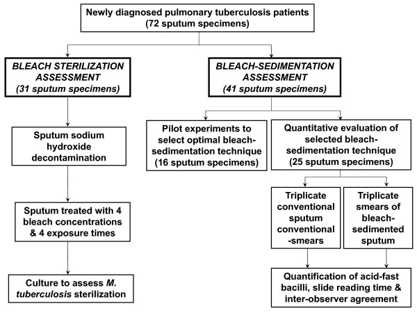
Methods: This blinded, controlled study used sputum specimens (n = 72) from tuberculosis patients. Bleach concentrations and exposure times required to sterilise sputum (n = 31) were determined. In the light of these results, the performance of 5 gravity bleach-sedimentation techniques that sterilise sputum specimens (n = 16) were compared. The best-performing of these bleach-sedimentation techniques involved adding 1 volume of 5% bleach to 1 volume of sputum, shaking for 10-minutes, diluting in 8 volumes distilled water and sedimenting overnight before microscopy. This technique was further evaluated by comparing numbers of visible acid-fast bacilli, slide-reading speed and reliability for triplicate smears before versus after bleach-sedimentation of sputum specimens (n = 25). Triplicate smears were made to increase precision and were stained using the Ziehl-Neelsen method.

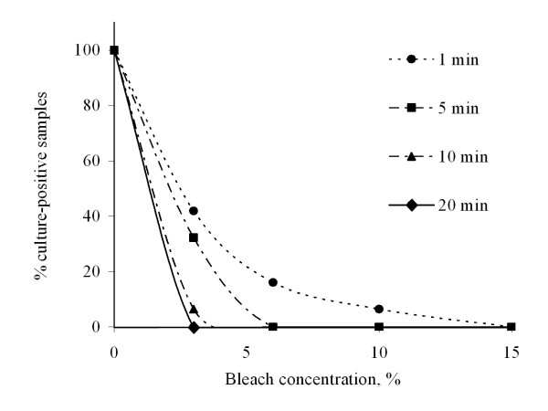
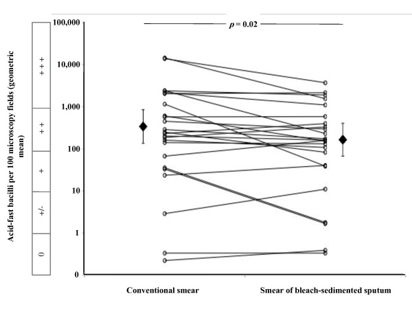
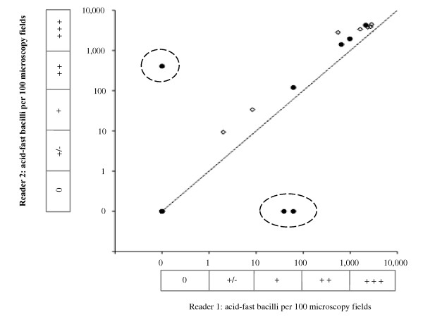
Results: M. tuberculosis in sputum was successfully sterilised by adding equal volumes of 15% bleach for 1-minute, 6% for 5-minutes or 3% for 20-minutes. Bleach-sedimentation significantly decreased the number of acid-fast bacilli visualised compared with conventional smears (geometric mean of acid-fast bacilli per 100 microscopy fields 166, 95%CI 68-406, versus 346, 95%CI 139-862, respectively; p = 0.02). Bleach-sedimentation diluted paucibacillary specimens less than specimens with higher concentrations of visible acid-fast bacilli (p = 0.02). Smears made from bleach-sedimented sputum were read more rapidly than conventional smears (9.6 versus 11.2 minutes, respectively, p = 0.03). Counting conventional acid-fast bacilli had high reliability (inter-observer agreement, r = 0.991) that was significantly reduced (p = 0.03) by bleach-sedimentation (to r = 0.707) because occasional strongly positive bleach-sedimented smears were misread as negative.

Conclusions: Gravity bleach-sedimentation improved laboratory safety by sterilising sputum but decreased the concentration of acid-fast bacilli visible on microscopy, especially for sputum specimens containing high concentrations of M. tuberculosis. Bleach-sedimentation allowed examination of more of each specimen in the time available but decreased the inter-observer reliability with which slides were read. Thus bleach-sedimentation effects vary depending upon specimen characteristics and whether microscopy was done for a specified time, or until a specified number of microscopy fields had been read. These findings provide an explanation for the contradictory results of previous studies.


Figures

Figure 1
Figure 1
Study flowchart. The Standards for the Reporting of Diagnostic Accuracy (STARD) flowchart for the study. Results were available for all of the procedures planned in the study protocol.
Figure 2
Figure 2
Bleach sterilization. M. tuberculosis viability after treatment of sputum specimens with 4 different bleach concentrations for 4 different exposure times, i.e. a total of 16 combinations of bleach concentrations and exposure times. All control specimens were culture-positive.
Figure 3
Figure 3
Effect of bleach-sedimentation on the concentration of acid-fast bacilli. The number of acid-fast bacilli visualised by smear-microscopy is shown. Each of the open circles represents the geometric mean number of acid-fast bacilli visible in 100 microscopy fields for triplicate, identically prepared slides. Each line joins the data derived from 1 of the 25 sputum specimens i.e. the geometric mean of triplicate conventional smear-microscopy slides (the left-hand end of each line) versus the geometric mean of triplicate slides prepared after bleach-sedimentation (the right-hand end of each line). The filled diamonds represent the geometric mean of all 25 specimens and the error bars represent 95% confidence intervals. The box parallel with the vertical axis indicates the smear-microscopy grade equivalent to the acid-fast bacilli counts per 100 microscopy fields (0 indicates none visible/100 fields; +/- indicates 1-9/100 fields; + indicates 10-99/100 fields; ++ indicates 100-999/100 fields; and +++ indicates > 1, 000/100 fields).
Figure 4
Figure 4
Correlation between the numbers of acid-fast bacilli visible by sputum smear-microscopy and the bleach-sedimentation concentrating effect. Each data point represents data from a single specimen. The horizontal axis shows the geometric mean number of acid-fast bacilli per 100 microscopy fields visualised in triplicate conventional smears. The horizontal error bars represent the standard error of the mean (SEM) for these triplicate data. The vertical axis shows the mean change in the number of acid-fast bacilli per 100 microscopy fields visualised in triplicate slides prepared after bleach-sedimentation of that specimen. The vertical error bars represent the SEM for these triplicate data. The diagonal broken line is the regression line. r represents the correlation coefficient. The box parallel with the horizontal axis indicates the smear-microscopy grade equivalent to the acid-fast bacilli counts per 100 microscopy fields (0 indicates none visible/100 fields; +/- indicates 1-9/100 fields; + indicates 10-99/100 fields; ++ indicates 100-999/100 fields; and +++ indicates > 1000/100 fields).
Figure 5
Figure 5
Microscopist inter-observer agreement. The inter-observer agreement is shown for 2 microscopists for conventional smears and for smears prepared after bleach-sedimentation. Filled circles represent smears prepared from bleach-sedimented sputum (correlation coefficient r = 0.707), and open diamonds represent conventional smears (r = 0.991). The 3 data points encircled by broken lines represent smears prepared from bleach-sedimented sputum found to be clearly positive by one reader but negative by the other reader. Excluding these 3 false-negative results caused the value of the correlation coefficient for results from bleach-sedimented sputum to increase to r = 0.997. The dotted line represents perfect agreement. The boxes parallel with the axes indicate the smear-microscopy grade equivalent to the acid-fast bacilli counts per 100 microscopy fields (0 indicates none visible/100 fields; +/- indicates 1-9/100 fields; + indicates 10-99/100 fields; ++ indicates 100-999/100 fields; and +++ indicates > 1000/100 fields).

References

    1. Nyirenda TE, Mundy CJ, Harries AD, Banerjee A, Salaniponi FM. Safety in laboratories carrying out sputum smear microscopy: a dilemma for resource-poor countries. Int J Tuberc Lung Dis. 1998;2:690–3. - PubMed
    1. Joshi R, Reingold AL, Menzies D, Pai M. Tuberculosis among health-care workers in low- and middle-income countries: a systematic review. PLoS Med. 2006;3:e494. doi: 10.1371/journal.pmed.0030494. - DOI - PMC - PubMed
    1. Kent PT, Kubica GP. Public health mycobacteriology: A guide for the level III laboratory. Atlanta: Centers for Disease Control; 1985.
    1. Best M, Sattar SA, Springthorpe VS, Kennedy ME. Efficacies of selected disinfectants against Mycobacterium tuberculosis. J Clin Microbiol. 1990;28:2234–2239. - PMC - PubMed
    1. Ängeby KAK, Alvarado-Galvez C, Pineda-Garcia L, Hoffner SE. Improved microscopy for a more sensitive diagnosis of pulmonary tuberculosis. Int J Tuberc Lung Dis. 2000;4:684–687. - PubMed

Publication types

MeSH terms