. 2011 Oct 10:11:59.
doi: 10.1186/1471-213X-11-59.

Genome-wide mapping of Sox6 binding sites in skeletal muscle reveals both direct and indirect regulation of muscle terminal differentiation by Sox6

Affiliations

Genome-wide mapping of Sox6 binding sites in skeletal muscle reveals both direct and indirect regulation of muscle terminal differentiation by Sox6

Chung-Il An et al. BMC Dev Biol. .

Abstract

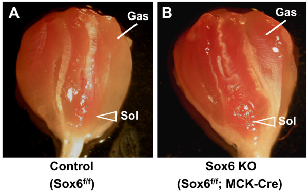
Background: Sox6 is a multi-faceted transcription factor involved in the terminal differentiation of many different cell types in vertebrates. It has been suggested that in mice as well as in zebrafish Sox6 plays a role in the terminal differentiation of skeletal muscle by suppressing transcription of slow fiber specific genes. In order to understand how Sox6 coordinately regulates the transcription of multiple fiber type specific genes during muscle development, we have performed ChIP-seq analyses to identify Sox6 target genes in mouse fetal myotubes and generated muscle-specific Sox6 knockout (KO) mice to determine the Sox6 null muscle phenotype in adult mice.

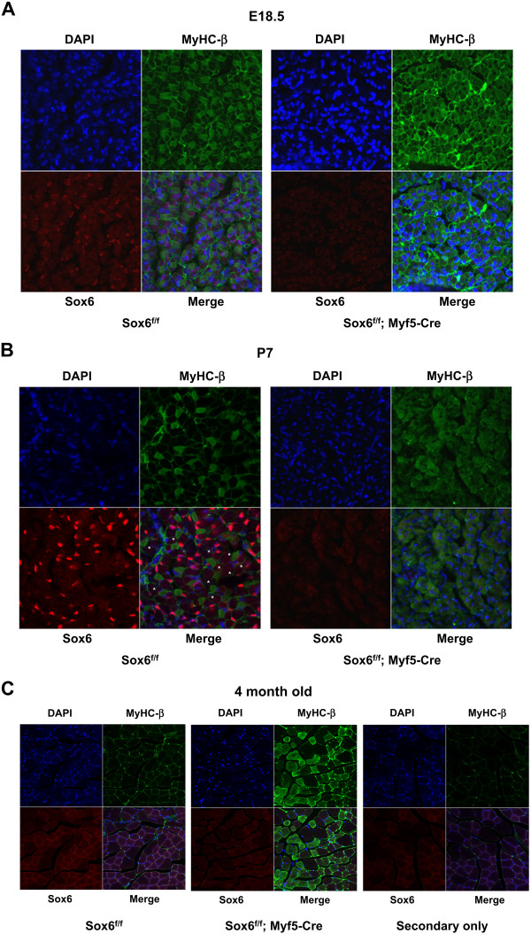
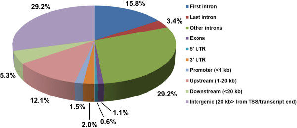
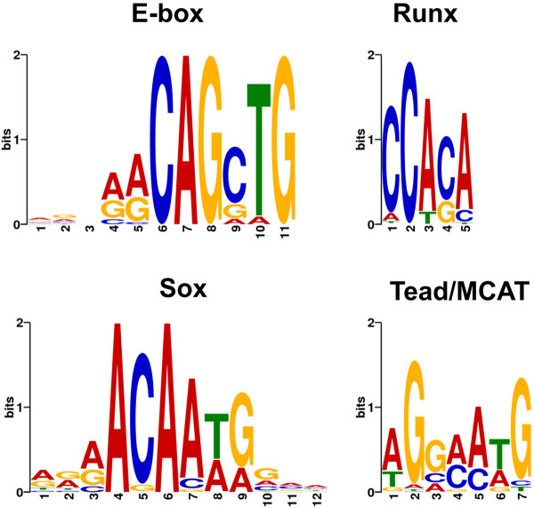
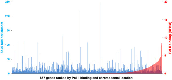
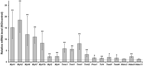
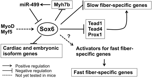
Results: We have identified 1,066 Sox6 binding sites using mouse fetal myotubes. The Sox6 binding sites were found to be associated with slow fiber-specific, cardiac, and embryonic isoform genes that are expressed in the sarcomere as well as transcription factor genes known to play roles in muscle development. The concurrently performed RNA polymerase II (Pol II) ChIP-seq analysis revealed that 84% of the Sox6 peak-associated genes exhibited little to no binding of Pol II, suggesting that the majority of the Sox6 target genes are transcriptionally inactive. These results indicate that Sox6 directly regulates terminal differentiation of muscle by affecting the expression of sarcomere protein genes as well as indirectly through influencing the expression of transcription factors relevant to muscle development. Gene expression profiling of Sox6 KO skeletal and cardiac muscle revealed a significant increase in the expression of the genes associated with Sox6 binding. In the absence of the Sox6 gene, there was dramatic upregulation of slow fiber-specific, cardiac, and embryonic isoform gene expression in Sox6 KO skeletal muscle and fetal isoform gene expression in Sox6 KO cardiac muscle, thus confirming the role Sox6 plays as a transcriptional suppressor in muscle development.

Conclusions: Our present data indicate that during development, Sox6 functions as a transcriptional suppressor of fiber type-specific and developmental isoform genes to promote functional specification of muscle which is critical for optimum muscle performance and health.


Figures

Figure 1
Figure 1
The number of fibers expressing MyHC-β is dramatically increased in Sox6 KO skeletal muscle. A. Cross-sections of E18.5 lower hindlimb muscle (TA-EDL region) from control (Sox6f/f) and Sox6KO (Sox6f/f; Myf5-Cre) were stained with DAPI (blue) or specific antibodies for MyHC-β (green) or Sox6 (red). x400 magnification. B. P7 (7 day old) hindlimb muscle processed for DAPI, Sox6 and MyHC-β immune-staining.* indicates myotubes that are negative for both MyHC-β and Sox6 staining in control muscle (see text for discussion). x400 magnification. C. Four month old adult TA-EDL muscle. A control muscle section stained with DAPI or secondary antibodies (a mixture of both anti-mouse and anti-rabbit) only was also shown. ×200 magnification.
Figure 2
Figure 2
Genome-wide mapping of Sox6 binding sites by ChIP-seq. Locations of Sox6 binding sites relative to the nearest RefSeq genes and the percentages of binding sites at the respective locations are shown.
Figure 3
Figure 3
Transcription factor consensus motifs found in Sox6 binding peaks.
Figure 4
Figure 4
Comparison of Sox6 binding and Pol II binding to the Sox6 target genes. Left Y axis shows fold enrichment of Sox6 obtained by the peak calling program SISSRs using the 3 million read data set (see the text for detail), and right Y axis shows Pol II binding levels to the Sox6 peak-associated genes measured in RPKM. X axis shows all Sox6-associated genes (867 RefSeq genes in total) sorted according to Pol II binding and chromosomal location. When multiple Sox6 peaks were associated with one gene, only the peak with the highest fold enrichment was used. Note that Pol II binding to β-actin, an abundantly expressed housekeeping gene, was 8.60 RPKM. A similar result was obtained using the 1.5 million read-data set (data not shown).
Figure 5
Figure 5
Differences of expression levels of Sox6 target genes between control and Sox6 KO perinatal mice. RT-qPCR was performed using total RNA from skeletal muscle of control (Sox6f/f) and Sox6 KO (Sox6f/f; MCK-Cre) newborn (postnatal day 1) mice. Expression levels in Sox6 KO mice were divided by those in control mice, and represented as mean ± SD (n = 3). The broken line corresponds to the expression ratio of 1, indicating equal expression level between the KO and control mice. (*) P < 0.05; (**) P < 0.005. Fiber type specific genes: Myh1, Myh2, Myh7, My7b, Myl2 (also expressed in the heart), Tnnc1 (also expressed in the heart), Tnni1, Tnnt1; cardiac isoforms: Myh6, Tnnt2; developmental isoform: Myl4; transcription factors: Prox1, Tcf4, Tead1, Tead4, Nfatc3; histone modification enzymes: Hdac9, Hdac11.
Figure 6
Figure 6
Morphological difference of skeletal muscle between control and Sox6 KO mice. Dissected gastrocnemius/soleus muscles from three month old mice are shown. A. control (Sox6f/f) muscle. B. Sox6 KO (Sox6f/f; MCK-Cre) muscle.
Figure 7
Figure 7
Nfatc3 protein levels are increased in both the nucleus and cytoplasm of Sox6-null myotubes. Wild type (WT) and Sox6-null (p100H) fetal primary myoblasts or myotubes were harvested at 0, 24, 48, 72, and 96 h after switching to DM, and nuclear and cytoplasmic protein was extracted for Nfatc3 Western blotting. TATA binding protein (TBP) and tubulin were used as loading controls for nuclear and cytoplasmic fractions, respectively. Upper panels: nuclear fractions. Lower panels: cytoplasmic fractions.
Figure 8
Figure 8
Differences of reporter activities between wild type and Sox6-null primary myotubes. A. Schematic representations of the firefly luciferase vectors constructed to test the function of Sox6 binding sequences. Black boxes indicate the firefly luciferase gene and shaded boxes indicate the chicken β-actin promoter (Actb-p). Open boxes indicate the upstream sequences of the wild type MyHC-β gene (MHCβ3500) or its mutated version (MHCβ3500 m; cross indicates mutation), Myh7b, Tnni1, and Hdac11, or the first intron sequence of Tnnc1. Approximate positions of Sox6 binding sites are indicated by gray lines. B. The reporter constructs shown in A were cotransfected with a Renilla luciferase vector into wild type (WT) and Sox6-null (p100H) primary myoblasts. After differentiation into myotubes, both luciferase activities were measured and normalized with Renilla luciferase activity. Data were further normalized to WT MHCβ3500 value (i.e. MHCβ3500 in WT = 1.0), and represented as mean ± SD (n = 3). (*) P < 0.05; (**) P < 0.005.
Figure 9
Figure 9
Summary of the present work concerning the fiber type specification under the control of Sox6. It has been shown that in zebrafish, MyoD and Myf5 are necessary to activate Sox6 gene expression in muscle [100]. This muscle-specific Sox6 activation mechanism has not been tested in mice yet, but since Sox6 upregulation coincides with upregulation of these myogenic regulatory factors during muscle differentiation, it is very likely that this mechanism is shared in mice also (see text). Once expressed in muscle, Sox6 directly suppresses transcription of slow fiber specific, cardiac and embryonic isoform genes during muscle development. In addition to these structural protein genes, Sox6 suppresses expression of the transcription factors which have been shown to activate slow fiber specific genes, Tead1, Tead4, and Prox1. By unknown mechanisms, fast fiber-specific gene expression in Sox6 KO skeletal muscle is dramatically reduced. Since Sox6 is preferentially expressed in fast-twitch fiber rich muscles, it is possible that Sox6 indirectly stimulates the fast fiber-specific gene program. This idea awaits future investigation. Sox6 activity in slow fibers is suppressed by miR-499 which is encoded in an intron of the Myh7b gene. We have shown that Sox6, in turn, suppresses Myh7b transcription. This negative feedback loop might be important for fiber type switching during muscle development as well as in adult muscle.

References

    1. Zierath JR, Hawley JA. Skeletal muscle fiber type: influence on contractile and metabolic properties. PLoS Biol. 2004;2:e348. doi: 10.1371/journal.pbio.0020348. - DOI - PMC - PubMed
    1. Pette D, Staron RS. Myosin isoforms, muscle fiber types, and transitions. Microsc Res Tech. 2000;50:500–9. doi: 10.1002/1097-0029(20000915)50:6<500::AID-JEMT7>3.0.CO;2-7. - DOI - PubMed
    1. Schiaffino S, Sandri M, Murgia M. Activity-dependent signaling pathways controlling muscle diversity and plasticity. Physiology (Bethesda) 2007;22:269–78. doi: 10.1152/physiol.00009.2007. - DOI - PubMed
    1. Gunning P, Hardeman E. Multiple mechanisms regulate muscle fiber diversity. FASEB J. 1991;5:3064–70. - PubMed
    1. Lu BD, Allen DL, Leinwand LA, Lyons GE. Spatial and temporal changes in myosin heavy chain gene expression in skeletal muscle development. Dev Biol. 1999;216:312–26. doi: 10.1006/dbio.1999.9488. - DOI - PubMed

Publication types

MeSH terms