Review
. 2012 Apr;24(4):528-38.
doi: 10.1111/j.1365-2826.2011.02236.x.

Cell-type specific expression of oxytocin and vasopressin genes: an experimental odyssey

Affiliations
Review

Cell-type specific expression of oxytocin and vasopressin genes: an experimental odyssey

H Gainer. J Neuroendocrinol. 2012 Apr.

Abstract

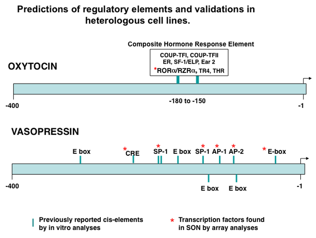
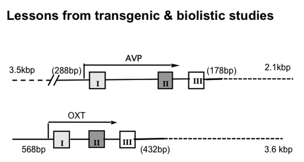
The supraoptic nucleus (SON) is a particularly good model for the study of cell-type specific gene expression because it contains two distinct neuronal phenotypes, the oxytocin (OT) and vasopressin (AVP) synthesising magnocellular neurones (MCNs). The MCNs are found in approximately equal numbers and selectively express either the OT or the AVP gene in approximately 97% of the MCN population in the SON. An unresolved issue has been to determine what mechanisms are responsible for the highly selective regulation of the cell-type specific expression of OT and AVP genes in the MCNs. Previous attempts to address this question have used various bioinformatic and molecular approaches, which included using heterologous cell lines to study the putative cis-elements in the OT and AVP genes, and the use of OT and/or AVP transgenes in transgenic rodents. The data from all of the above studies identified a region < 0.6 kbp upstream of OT exon I and approximately 3 kb upstream of AVP exon I as being sufficient to produce cell-specific expression of the OT and AVP genes, respectively, although they failed to identify the specific cis-domains responsible for the MCN-specific gene expression. An alternative experimental approach to perform promoter deletion analysis in vivo (i.e. to use stereotaxic viral vector gene transfer into the SON to further dissect the cis-elements in the OT and AVP genes) will be described here. This in vivo method uses adeno-associated viral (AAV) vectors expressing OT-promoter deletion constructs and utilises the enhanced green fluorescent protein (EGFP) as the reporter. The AAV constructs are stereotaxically injected into the rat brain above the SON and, 2 weeks post injection, the rats are sacrificed and assayed for EGFP expression. Using this method, it has been possible to identify specific regions upstream of the transcription start site in the OT and AVP gene promoters that are responsible for conferring the cell-type specificity of the OT and AVP gene expression in the SON.


Figures

Figure 1
Figure 1
Magnocellular Neuronal Phenotypes in the rat SON. A. Shows double-label immunofluorescent images for oxytocin (green) and vasopressin (red) containing magnocellular neurons (MCNs) in the rat SON. These two distinct MCN populations are the only neuronal phenotypes found in the SON. Most of the MCNs express only oxytocin or vasopressin, however, a small percentage of the MCNs (about 2–3%) are known to express both peptides (see arrows). Scale bar = 100µm. Abbreviations: OC, optic chiasm. B. Oxytocin and vasopressin phenotypes of individual dissociated hypothalamic supraoptic magnocellular neurons were determined by RT-PCR. A photomicrograph is shown of an individual dissociated magnocellular neuron being picked up by a harvesting pipette touching the large cell body containing two large dendrites. Scale bar, 20µm. cDNAs from the individual cells were amplified by PCR with primers that are specific for oxytocin (OT), vasopressin (VP), or glyceraldehyde 3-phosphate dehydrogenase (GAPDH). The PCR products were then run on 1.5% agarose gels containing ethidium bromide.
Figure 2
Figure 2
Regulatory elements in the OXT and AVP promoters 500bp upstream of their transcription start sites that are predicted by bioinformatic analyses and which were functionally validated by experiments using heterologous cell lines (based on data described in Burbach et al, (3). The asterisks show the transcription factors that bind to these elements which were detected in the SON by microarray analyses (based on data in Hindmarch(82); Mutsuga et. al, (83); and Yue et. al, (84)). Abbreviations: COUP-TF I and II:Chicken ovalbumin upstream promoter transcription factors I and II; ER: estrogen receptor; SF-1/ELP: steroidogenic factor; Ear2: V-erb-related protein 2 transcription factor; RORa/RZRa: retinoic acid receptor orphan receptor transcription factor family; THR: thyroid hormone receptor; CRE: camp response element; SP1: specificity protein 1 transcription factor; AP1: activator protein 1; AP2: activator protein 2, Ebox: DNA sequence, CANNTG/CACGTG,that binds helix loop-helix transcription factors (e.g., BMAL-CLOCK).
Figure 3
Figure 3
Summary of results from Transgenic and Organotypic Culture/Biolistics Studies (1987–2003). These constructs were found to produce cell-type specific expression in the Oxt and Avp MCNs in the SON. In transgenic rodents the Avp gene with 3.5kbp upstream and 2.1kbp downstream (dotted line) of the gene body produced cell-type specific expression, whereas using biolistics and organotypic culture only 288bp upstream and 178bp downstream were found to be neccessary. For the Oxt gene 568bp upstream and 3.6kbp downstream of the gene body in the transgene produced cell-type specific expression, whereas in biolistic experiments 568bp upstream and only 432bp downstream were found to be sufficient (see reviews by Young and Gainer, (10); and Murphy and Wells(11)).
Figure 4
Figure 4
Use of Adeno-Associated Virus (AAV) to study gene expression in MCNs in organotypic hypothalamic cultures. Top: Genomic organization of AAV. The AAV genome produces four transcripts, Rep 40,52, 68 and 78 which are genes involved in replication, and three proteins, VP-1,-2 and-3 which are genes for forming the capsid. The ITRs (Inverted Terminal Repeat Sequences) are necessary for viral replication, rescue, packaging and integration. Bottom: AAV transduction of hypothalamic slice explant cultures. Slices were incubated in AAV-CMV-nLac-Z for 7 days and following this incubation, the tissues were fixed in 2 % paraformaldehyde and double immunostained for nuclear localized lac-z immunoreactivity (black) and oxytocin-neurophysin immunoreactivity (brown). A and B: Control slices (no AAV added). C and D: AAV transduced slices.(adapted from Kier et. al., (66)). The DAB (brown) stained OXT neurons shown in B and D are in the Accessory Nucleus in the hypothalamic slice.
Figure 5
Figure 5
Use of Adeno-Associated Virus (AAV) to study cell-type specific expression in (A) Avp and (B) Oxt MCNs in vivo. Stereotaxic injections of rAAV vectors containing construct with either Avp (in A) or Oxt (in B) promoters fused to EGFP reporters produced cell-specific expression of the EGFP in the injected SONs. A. Shows EGFP expression in the MCNs, which co-localize with Avp-immunoreactivity (Avp-ir), but not with Oxt-immunoreactivity (Oxt-ir). B. Shows EGFP expression in the MCNs, which co-localize with Oxt-ir, but not with Avp-ir. The IGR is the intergenic region sequence as defined in Fields et. al.(2003; ref 50). For the AVP construct the IGR was 181bp, for the OXT construct the IGR was 442bp. The ITRs (Inverted Terminal Repeat Sequences) are necessary for viral replication, rescue, packaging and integration (see Fig. 4 top).
Figure 6
Figure 6
Comparisons of cis-domains in the Avp and Oxt gene promoters that are found to be important for their cell type specific expression before and after application of the AAV strategy.

Comment in

Similar articles

Cited by

References

    1. Armstrong WE. Morphological and electrophysiological classification of hypothalamic supraoptic neurons. Prog Neurobiol. 1995;47(4–5):291–339. - PubMed
    1. Hatton GI. Oxytocin and vasopressin neurones: vive la difference! J Physiol. 1997;500(Pt 2):284. - PMC - PubMed
    1. Burbach JP, Luckman SM, Murphy D, Gainer H. Gene regulation in the magnocellular hypothalamo-neurohypophysial system. Physiol Rev. 2001;81(3):1197–1267. - PubMed
    1. Vandesande F, Dierickx K. Identification of the vasopressin producing and of the oxytocin producing neurons in the hypothalamic magnocellular neurosecretroy system of the rat. Cell Tissue Res. 1975;164(2):153–162. - PubMed
    1. Mohr E, Bahnsen U, Kiessling C, Richter D. Expression of the vasopressin and oxytocin genes in rats occurs in mutually exclusive sets of hypothalamic neurons. FEBS Lett. 1988;242(1):144–148. - PubMed

Publication types

MeSH terms