. 2011 Nov;121(11):4409-19.
doi: 10.1172/JCI58097. Epub 2011 Oct 10.

Parabronchial smooth muscle constitutes an airway epithelial stem cell niche in the mouse lung after injury

Affiliations

Parabronchial smooth muscle constitutes an airway epithelial stem cell niche in the mouse lung after injury

Thomas Volckaert et al. J Clin Invest. 2011 Nov.

Abstract

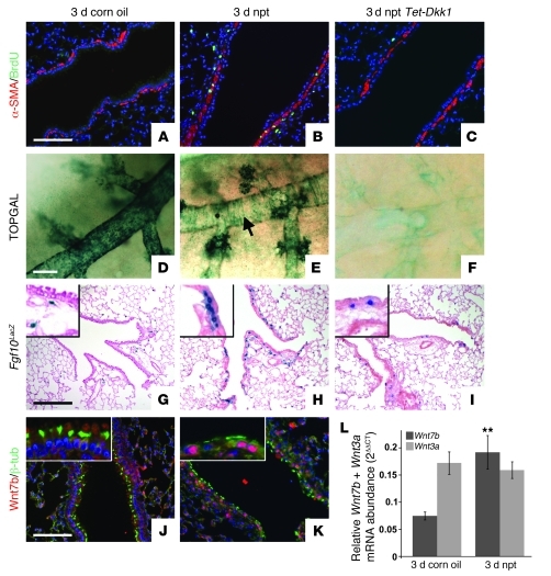
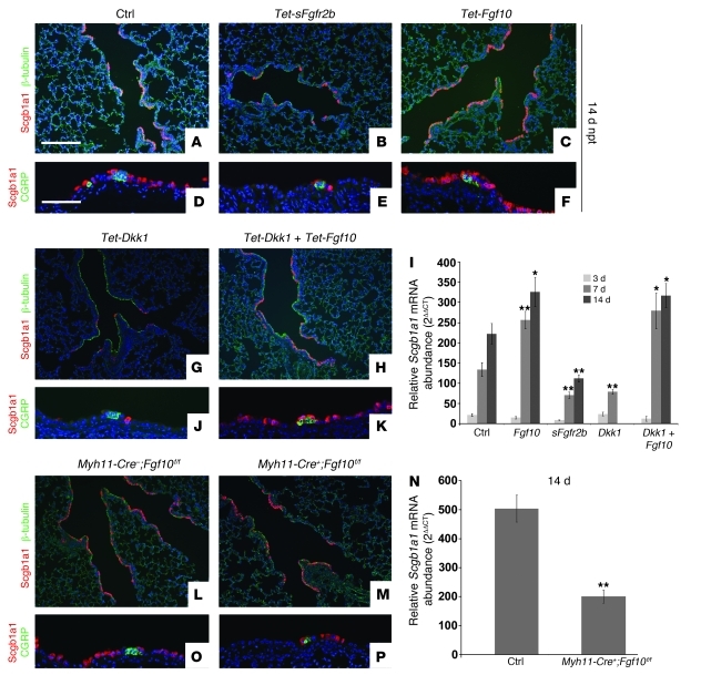
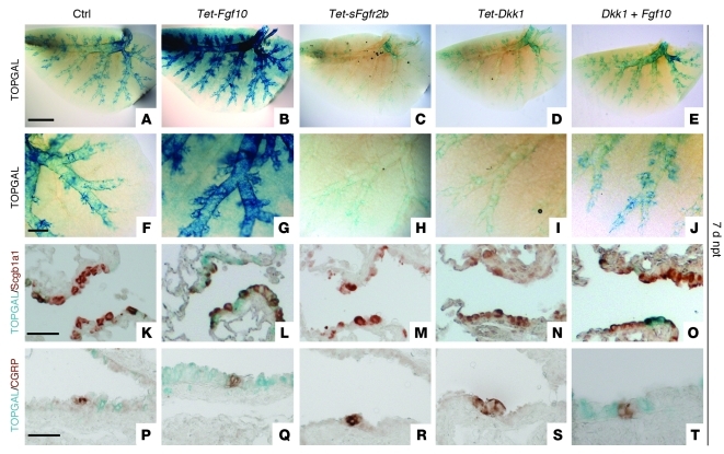
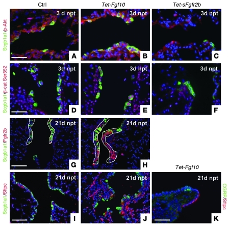
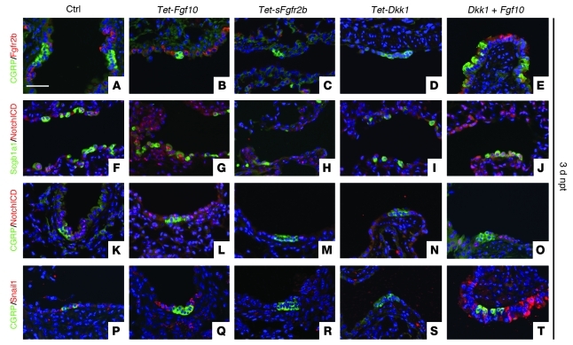
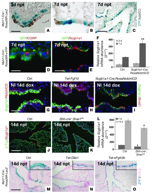
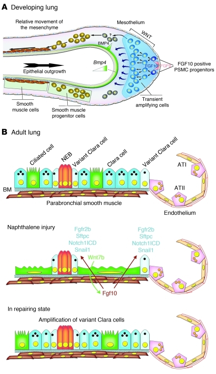
During lung development, parabronchial SMC (PSMC) progenitors in the distal mesenchyme secrete fibroblast growth factor 10 (Fgf10), which acts on distal epithelial progenitors to promote their proliferation. β-catenin signaling within PSMC progenitors is essential for their maintenance, proliferation, and expression of Fgf10. Here, we report that this Wnt/Fgf10 embryonic signaling cascade is reactivated in mature PSMCs after naphthalene-induced injury to airway epithelium. Furthermore, we found that this paracrine Fgf10 action was essential for activating surviving variant Clara cells (the cells in the airway epithelium from which replacement epithelial cells originate) located at the bronchoalveolar duct junctions and adjacent to neuroendocrine bodies. After naphthalene injury, PSMCs secreted Fgf10 to activate Notch signaling and induce Snai1 expression in surviving variant Clara cells, which subsequently underwent a transient epithelial to mesenchymal transition to initiate the repair process. Epithelial Snai1 expression was important for regeneration after injury. We have therefore identified PSMCs as a stem cell niche for the variant Clara cells in the lung and established that paracrine Fgf10 signaling from the niche is critical for epithelial repair after naphthalene injury. These findings also have implications for understanding the misregulation of lung repair in asthma and cancer.


Figures

Figure 1
Figure 1. Wnt7b expressed by surviving ciliated cells induces Fgf10 expression in PSMCs 3 days after naphthalene-mediated Clara cell injury.
(AC) Immunostaining for proliferation marker BrdU and SMC marker α-SMA on 2-month-old WT lungs 3 days after corn oil treatment (A), WT lungs 3 days after naphthalene (npt) treatment (B), and Rosa26-rtTa;Tet-Dkk1 lungs 3 days after naphthalene treatment (C). (DF) β-gal staining on 2-month-old TOPGAL lungs 3 days after corn oil treatment (D), TOPGAL lungs 3 days after naphthalene treatment (E), and Rosa26-rtTa;Tet-Dkk1;TOPGAL lungs 3 days after naphthalene treatment (F). Arrow in E denotes TOPGAL activation in PSMCs. (GI) β-gal staining on 2-month-old Fgf10LacZ lungs 3 days after corn oil treatment (G), Fgf10LacZ lungs 3 days after naphthalene treatment (H), and Rosa26-rtTa;Tet-Dkk1;Fgf10LacZ lungs 3 days after naphthalene treatment (I). Insets are enlarged ×4. Note that we identified the blue cells in alveolar compartment as lipofibroblasts (S.P. De Langhe, unpublished observations). (J and K) Immunostaining for ciliated cell marker β-tubulin and Wnt7b on 2-month-old WT lungs 3 days after corn oil treatment (J) or naphthalene treatment (K). Insets are enlarged ×3. (L) qPCR analysis of relative Wnt7b and Wnt3a mRNA abundance in 2-month-old WT lungs 3 days after treatment with corn oil versus naphthalene. **P < 0.01 vs. respective control. n = 3. Scale bars: 100 μm (AC, J, and K); 250 μm (DF); 200 μm (GI).
Figure 2
Figure 2. Wnt-induced Fgf10 secreted by PSMCs is essential for epithelial repair after naphthalene injury.
(AC, G, H, L, and M) Immunostaining for Clara cell marker Scgb1a1 and ciliated cell marker β-tubulin on lungs 14 days after naphthalene treatment isolated from control (A), dox-induced Rosa26-rtTa;Tet-sFgfr2b (B), dox-induced Rosa26-rtTA;Tet-Fgf10 (C), dox-induced Rosa26-rtTa;Tet-Dkk1 (G), dox-induced Rosa26-rtTa;Tet-Dkk1;Tet-Fgf10 (H), Myh11-Cre;Fgf10fl/fl (L), and Myh11-Cre+;Fgf10fl/fl (M) mice. (DF, J, K, O, and P) Immunostaining for Scgb1a1 and neuroendocrine marker CGRP on lungs 14 days after naphthalene treatment isolated from control (D), dox-induced Rosa26-rtTa;Tet-sFgfr2b (E), dox-induced Rosa26-rtTA;Tet-Fgf10 (F), dox-induced Rosa26-rtTa;Tet-Dkk1 (J), dox-induced Rosa26-rtTa;Tet-Dkk1;Tet-Fgf10 (K), Myh11-Cre;Fgf10fl/fl (O), and Myh11-Cre+;Fgf10fl/fl (P) mice. (I) qPCR analysis of relative Scgb1a1 mRNA abundance of adult lungs from control, Rosa26-rtTa;Tet-sFgfr2b, Rosa26-rtTA-Tet-Fgf10, Rosa26rtTa;Tet-Dkk1, and Rosa26-rtTa;Tet-Dkk1;Tet-Fgf10 mice 3, 7, and 14 days after naphthalene treatment. (N) qPCR analysis of relative Scgb1a1 mRNA abundance in lungs from 2-month-old Myh11-Cre;Fgf10fl/fl and Myh11-Cre+;Fgf10fl/fl mice 14 days after naphthalene treatment. **P < 0.01, *P < 0.05 vs. respective control. n ≥ 3. Scale bars: 200 μm (AC, G, H, L, and M); 100 μm (DF, J, K, O, and P).
Figure 3
Figure 3. Fgf10 mediates epithelial repair independent of Wnt ligands in part by activating/enhancing β-catenin signaling in the epithelium.
β-gal staining (AJ) and β-gal staining with coimmunostaining for Scgb1a1 (KO) or CGRP (PT) on lungs 7 days after naphthalene treatment from TOPGAL (A, F, K, and P), dox-induced Rosa26-rtTA;Tet-Fgf10;TOPGAL (B, G, L, and Q), dox-induced Rosa26-rtTa;Tet-sFgfr2b;TOPGAL (C, H, M, and R), dox-induced Rosa26-rtTa;Tet-Dkk1;TOPGAL (D, I, N, and S), and dox-induced Rosa26-rtTa;Tet-Dkk1;Tet-Fgf10;TOPGAL mice (E, J, O, and T). Scale bars: 2,000 μm (AE); 500 μm (FJ); 50 μm (KT).
Figure 4
Figure 4. Fgf10 signaling induces Akt-mediated phosphorylation of β-catenin and maintenance/amplification of variant Clara cells.
(AC) Immuno­staining for p-Akt and Scgb1a1 on lungs from control (A), Rosa26-rtTa;Tet-Fgf10 (B), and Rosa26-rtTA;Tet-sFgfr2b (C) mice 3 days after naphthalene treatment. (DF) Immuno­staining for p–β-catenin–Ser552 and Scgb1a1 on lungs from control (D), Rosa26-rtTa;Tet-Fgf10 (E), and Rosa26-rtTA;Tet-sFgfr2b (F) mice 3 days after naphthalene treatment. (G and H) Immunostaining for Scgb1a1 and Fgfr2b on lungs from control (G) and Rosa26-rtTa;Tet-Fgf10 (H) mice 21 days after naphthalene treatment. White outlines denote the epithelium at the BADJ. (I and J) Immuno­staining for BASC markers Scgb1a1 and Sftpc on lungs from control (I) and Rosa26-rtTa;Tet-Fgf10 (J) mice 21 days after naphthalene treatment. (K) Immuno­staining for CGRP and Sftpc on lungs from Rosa26-rtTa;Tet-Fgf10 mice 21 days after naphthalene treatment. Scale bars: 50 μm (AF and K); 100 μm (GJ).
Figure 5
Figure 5. Flow cytometry analysis of airway epithelial stem cells.
Flow cytometry analysis using Sftpc and Scgb1a1 (A, C, and E) versus Fgfr2 and Scgb1a1 (B, D, and F) antibodies on permeabilized epithelial cells from WT noninjured lungs (A and B), WT lungs 7 days after naphthalene injury (C and D), and Rosa26-rtTa;Tet-Fgf10 lungs 7 days after naphthalene injury (E and F). A 14-fold increase in double-positive airway epithelial cells was detected with both antibody combinations 7 days after naphthalene injury; a 22- to 33-fold increase was seen 7 days after injury in mice overexpressing Fgf10. Forward scatter/side scatter (FS/SS) plots indicated that the same pool of airway epithelial stem cells was isolated using either antibody combination.
Figure 6
Figure 6. Fgf10 signaling induces Fgfr2b expression, Notch activation, and subsequent Snail induction in the activated variant Clara cells.
Immunostaining for CGRP and Fgfr2b (AE), Scgb1a1 and NotchICD (active notch) (FJ), CGRP and NotchICD (KO), and CGRP and Snail1 (PT) on lungs from control (A, F, K, and P), dox-induced Rosa26-rtTa;Tet-Fgf10 (B, G, L, and Q), dox-induced Rosa26-rtTA;Tet-sFgfr2b (C, H, M, and R), dox-induced Rosa26-rtTa;Tet-Dkk1 (D, I, N, and S), and dox-induced Rosa26-rtTa;Tet-Dkk1;Tet-Fgf10 (E, J, O, and T) mice 3 days after naphthalene treatment. Scale bar: 50 μm (AT).
Figure 7
Figure 7. Variant Clara cells undergo transient EMT in response to Fgf10-induced Notch activation and subsequent Snail induction.
(AC) Lineage tracing of activated variant Clara cells that underwent transient EMT in Myh11-Cre;Rosa26-LacZ mice 3 (A) and 7 (B and C) days after naphthalene injury. (C) Lower-magnification view of B. (D and E) Lineage tracing of activated variant Clara cells that underwent transient EMT in Myh11-Cre;Rosa26-eYFP mice 7 days after naphthalene injury, shown by immunostaining for GFP and CGRP (D) or GFP and Scgb1a1 (E). (F) qPCR analysis of relative Scgb1a1 mRNA abundance in lungs from 2-month-old control and Scgb1a1-rtTa;Tet-o-Cre;Rosa26-NotchICD mice 3 and 7 days after naphthalene treatment. (G and H) Immunostaining for NotchICD and Scgb1a1 on noninjured (NI) lungs from control (G) and Rosa26-rtTa;Tet-Fgf10 mice (H) 14 days after dox treatment. (I) Immunostaining for Snail1 and Scgb1a1 on noninjured lungs from Scgb1a1-rtTa;Tet-o-Cre;Rosa26-NotchICD+/– mice 14 days after dox treatment. (J and K) Immunostaining for Scgb1a1 and β-tubulin on lungs from control (J) and Shh-Cre+;Snai1fl/fl (K) mice 14 days after naphthalene treatment. (L) qPCR analysis of relative Scgb1a1 mRNA abundance in lungs from 2-month-old control and Shh-Cre+;Snai1fl/fl mice 7 and 14 days after naphthalene treatment. (MO) Lineage tracing of activated variant Clara cells that underwent transient EMT in Myh11-Cre;Rosa26-LacZ (M), Myh11-Cre;Rosa26-LacZ;Rosa26-rtTa;Tet-Dkk1 (N), and Myh11-Cre;Rosa26-LacZ;Rosa26-rtTA;Tet-sFgfr2b (O) mice 14 days after naphthalene injury. Insets are enlarged ×4. **P < 0.01, *P < 0.05 vs. control. n ≥ 3. Scale bars: 50 μm (A, B, E, and GI); 200 μm (C, J, K, and MO); 31.7 μm (D).
Figure 8
Figure 8. Model of PSMC progenitors during development and recapitulation of a progenitor state by mature PSMCs after naphthalene injury.
(A) During embryonic lung development, PSMC progenitors are identified as Fgf10-expressing cells in the distal mesenchyme (10). The amplification of these PSMC progenitors as well as their Fgf10 expression is regulated by mesenchymal β-catenin signaling (9). Fgf10 signaling is critical during lung development in the maintenance of lung epithelial progenitors (5, 8, 13). (B) In the adult lung, Fgf10 expression in mature PSMCs is silenced. Upon naphthalene administration, Clara cells are destroyed — except for variant Clara cells — at both NEBs and BADJs. Ciliated cells spread out to cover the basement membrane (BM) and induce Wnt7b expression. Wnt7b then acts on the PSMCs to reactivate Fgf10 expression, which then acts on the variant Clara cells to activate Notch signaling and induce expression of Fgfr2b, Sftpc, and Snail1, thereby initiating the repair process through a transient EMT. ATI, alveolar type I cell; ATII, alveolar type II cell; NEB, neuroendocrine body.

References

    1. Giangreco A, Reynolds SD, Stripp BR. Terminal bronchioles harbor a unique airway stem cell population that localizes to the bronchoalveolar duct junction. Am J Pathol. 2002;161(1):173–182. doi: 10.1016/S0002-9440(10)64169-7. - DOI - PMC - PubMed
    1. Reynolds SD, Giangreco A, Power JH, Stripp BR. Neuroepithelial bodies of pulmonary airways serve as a reservoir of progenitor cells capable of epithelial regeneration. Am J Pathol. 2000;156(1):269–278. doi: 10.1016/S0002-9440(10)64727-X. - DOI - PMC - PubMed
    1. Chen H, Matsumoto K, Stripp BR. Bronchiolar progenitor cells. Proc Am Thorac Soc. 2009;6(7):602–606. doi: 10.1513/pats.200907-078RM. - DOI - PMC - PubMed
    1. Rawlins EL, Hogan BL. Epithelial stem cells of the lung: privileged few or opportunities for many? Development. 2006;133(13):2455–2465. doi: 10.1242/dev.02407. - DOI - PubMed
    1. Bellusci S, Grindley J, Emoto H, Itoh N, Hogan BL. Fibroblast growth factor 10 (FGF10) and branching morphogenesis in the embryonic mouse lung. Development. 1997;124(23):4867. - PubMed

Publication types

MeSH terms