. 2011 Dec;179(6):2885-93.
doi: 10.1016/j.ajpath.2011.08.030. Epub 2011 Oct 8.

Starvation compromises Paneth cells

Affiliations

Starvation compromises Paneth cells

Caroline M Hodin et al. Am J Pathol. 2011 Dec.

Abstract

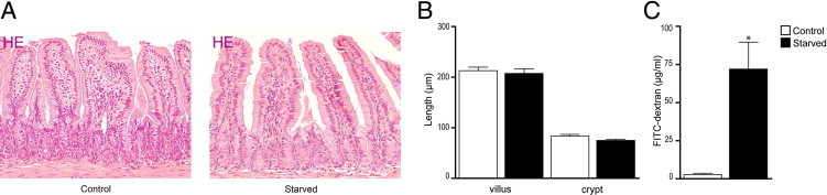
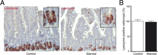
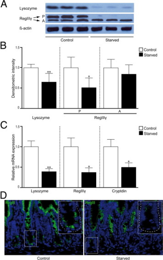
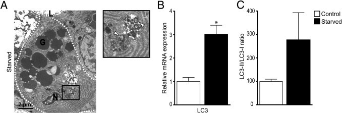
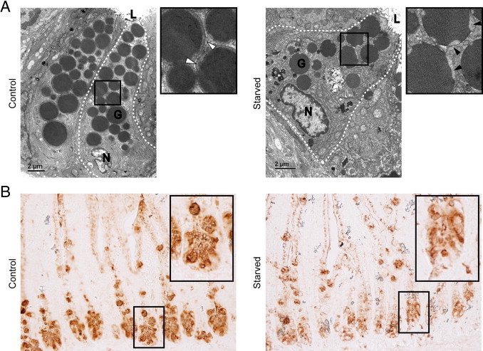
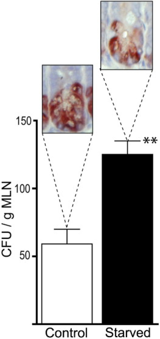
Lack of enteral feeding, with or without parenteral nutritional support, is associated with increased intestinal permeability and translocation of bacteria. Such translocation is thought to be important in the high morbidity and mortality rates of patients who receive nothing by mouth. Recently, Paneth cells, important constituents of innate intestinal immunity, were found to be crucial in host protection against invasion of both commensal and pathogenic bacteria. This study investigates the influence of food deprivation on Paneth cell function in a mouse starvation model. Quantitative PCR showed significant decreases in mRNA expression of typical Paneth cell antimicrobials, lysozyme, cryptdin, and RegIIIγ, in ileal tissue after 48 hours of food deprivation. Protein expression levels of lysozyme and RegIIIγ precursor were also significantly diminished, as shown by Western blot analysis and IHC. Late degenerative autophagolysosomes and aberrant Paneth cell granules in starved mice were evident by electron microscopy, Western blot analysis, and quantitative PCR. Furthermore, increased bacterial translocation to mesenteric lymph nodes coincided with Paneth cell abnormalities. The current study demonstrates the occurrence of Paneth cell abnormalities during enteral starvation. Such changes may contribute to loss of epithelial barrier function, causing the apparent bacterial translocation in enteral starvation.


Figures

Figure 1
Figure 1
Morphological and functional changes in the starved intestine. A: H&E staining of ileum from control (left panel) and starved (right panel) mice showed no apparent changes in intestinal architecture. B: Villus length and crypt depth were similar in the ileum of starved mice compared with controls. C: Assessment of ileal permeability demonstrated a significant increase in plasma 4.4-kDa FITC-dextran of starved mice compared with controls. *P < 0.05. The histological features are representative for all tissue samples studied (n = 15 per group).
Figure 2
Figure 2
Starvation results in decreased lysozyme expression in Paneth cells. A: IHC for lysozyme in ileum from control (left panel) versus starved (right panel) mice showed reduced expression of lysozyme in Paneth cells from fasted animals. Insets: Magnifications of single crypts from both control and starved mice. B: The number of Paneth cells per crypt in the ileum of starved and control mice showed no difference. The histological features are representative for all tissue samples studied (n = 15 per group).
Figure 3
Figure 3
Protein and mRNA levels of Paneth cell antimicrobial proteins are decreased after 48 hours of starvation. A: Western blot analysis showed decreased band density for lysozyme (15 kDa) and the precursor (P) form of RegIIIγ (16.5 kDa) in the ileum of starved mice compared with control mice, whereas no change was observed in the active form (A) of RegIIIγ (representative bands of three mice per group are shown). β-Actin was used to correct for equal protein loading (n = 12 per group). B: Quantification of Western blots demonstrated a significant decrease in both lysozyme (*P < 0.01) expression and expression of the precursor form of RegIIIγ (**P < 0.05). C: qPCR analysis showed a 2.5-fold decrease in lysozyme gene expression (*P < 0.01), a 2.7-fold decrease in gene expression of RegIIIγ (**P < 0.05), and a twofold decrease in cryptdin (**P < 0.05) gene expression after starvation (n = 15 per group). D: Immunofluorescence for RegIIIγ in ileum from control (left panel) versus starved (right panel) mice showed an overall reduced expression of RegIIIγ in fasted animals, as well as in Paneth cells specifically. Insets: Magnifications of single crypts from both control and starved mice. The histological features are representative for all tissue samples studied (n = 15 per group).
Figure 4
Figure 4
Autophagy in Paneth cells of starved mice. A: Electron microscopy showed the presence of late degradative autophagosomes (white arrowheads). G, granule; L, lumen; N, nucleus. B: qPCR showed a significant increase in LC3 gene expression in the small intestine of starved mice. *P < 0.01. C: Quantification of band density of phosphatidylethanolamine-conjugated LC3 (LC3-II) and LC3-I, as assayed by using Western blot analysis, revealed a trend for an increase in the LC3-II/LC-I ratio. P = 0.10.
Figure 5
Figure 5
Granule morphological features and composition is aberrant in Paneth cells of starved mice. A: Electron microscopy revealed changes in the electron-lucent halo of granules in Paneth cells of starved mice (right panel, magnification, black arrowheads) compared with control mice (left panel, magnification, white arrowheads). The dashed lines demarcate Paneth cells. G, granule; L, lumen; N, nucleus (n = 3 per group). B: Staining with lectin T. vulgaris showed less intense staining and aberrant granule morphological features in Paneth cells of starved mice (n = 15 per group). Images are representative for all tissue samples studied.
Figure 6
Figure 6
Enhanced bacterial translocation to the MLNs in starved mice. Bacterial translocation to MLNs is significantly elevated in mice with decreased lysozyme staining after 48 hours of starvation compared with controls [59.1 ± 10.8 versus 125.1 ± 9.9 colony-forming units per gram (CFU/g) of MLNs]. **P < 0.01 (n = 10 per group).

References

    1. Bevins C.L., Salzman N.H. Paneth cells, antimicrobial peptides and maintenance of intestinal homeostasis. Nat Rev Microbiol. 2011;9:356–368. - PubMed
    1. Ayabe T., Satchell D.P., Wilson C.L., Parks W.C., Selsted M.E., Ouellette A.J. Secretion of microbicidal alpha-defensins by intestinal Paneth cells in response to bacteria. Nat Immunol. 2000;1:113–118. - PubMed
    1. Vaishnava S., Behrendt C.L., Ismail A.S., Eckmann L., Hooper L.V. Paneth cells directly sense gut commensals and maintain homeostasis at the intestinal host-microbial interface. Proc Natl Acad Sci U S A. 2008;105:20858–20863. - PMC - PubMed
    1. Grootjans J., Hodin C.M., de Haan J.J., Derikx J.P., Rouschop K.M., Verheyen F.K., van Dam R.M., Dejong C.H., Buurman W.A., Lenaerts K. Level of activation of the unfolded protein response correlates with Paneth cell apoptosis in human small intestine exposed to ischemia/reperfusion. Gastroenterology. 2011;140:529–539. - PubMed
    1. MacFie J. Enteral versus parenteral nutrition: the significance of bacterial translocation and gut-barrier function. Nutrition. 2000;16:606–611. - PubMed

Publication types

MeSH terms