. 2012 Apr;22(4):746-56.
doi: 10.1038/cr.2011.162. Epub 2011 Oct 11.

Revealing a steroid receptor ligand as a unique PPARγ agonist

Affiliations

Revealing a steroid receptor ligand as a unique PPARγ agonist

Shengchen Lin et al. Cell Res. 2012 Apr.

Abstract

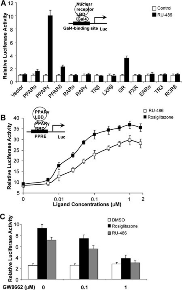
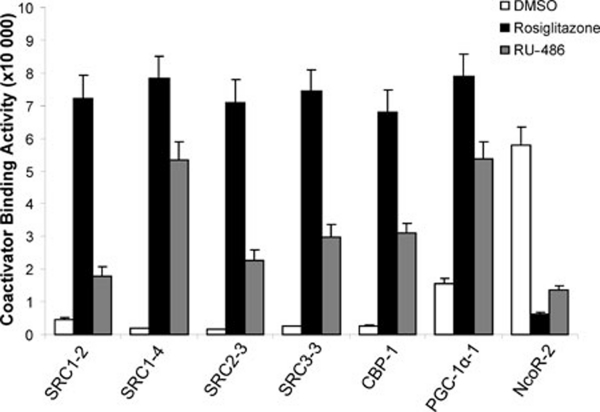
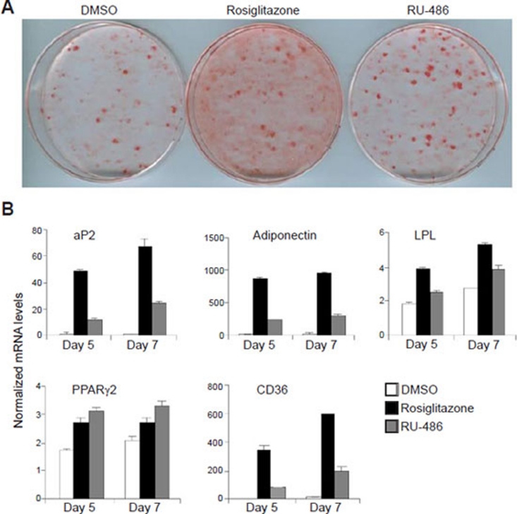
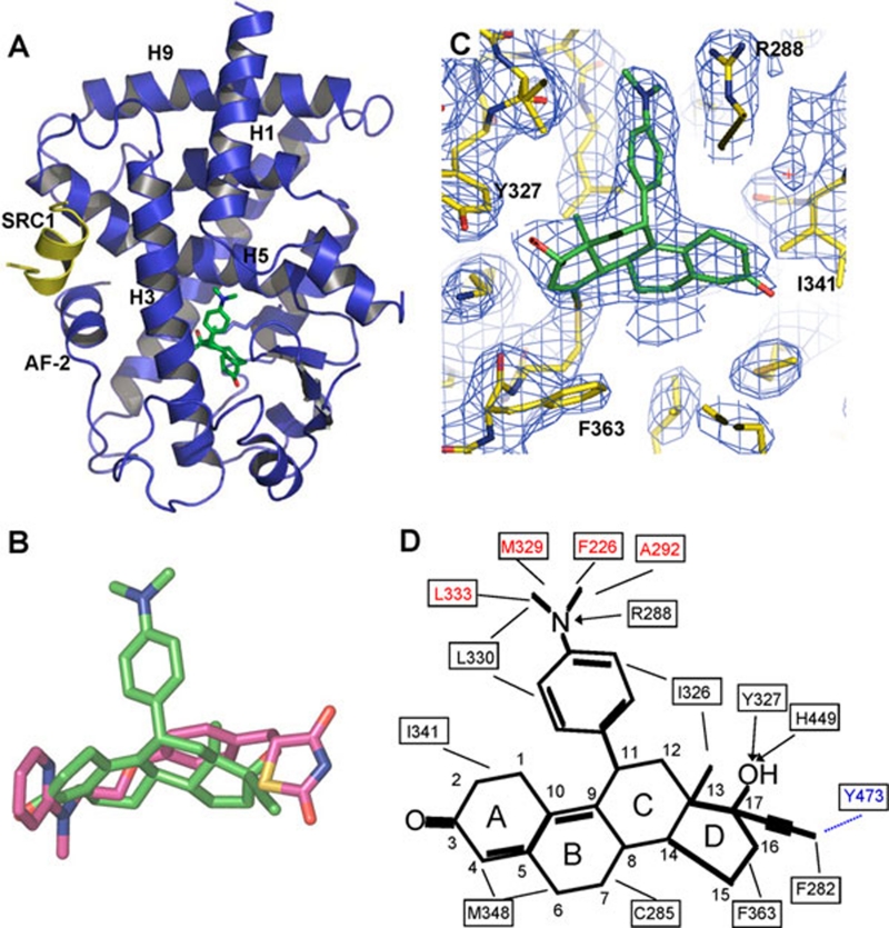
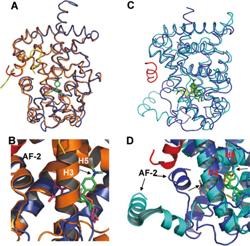
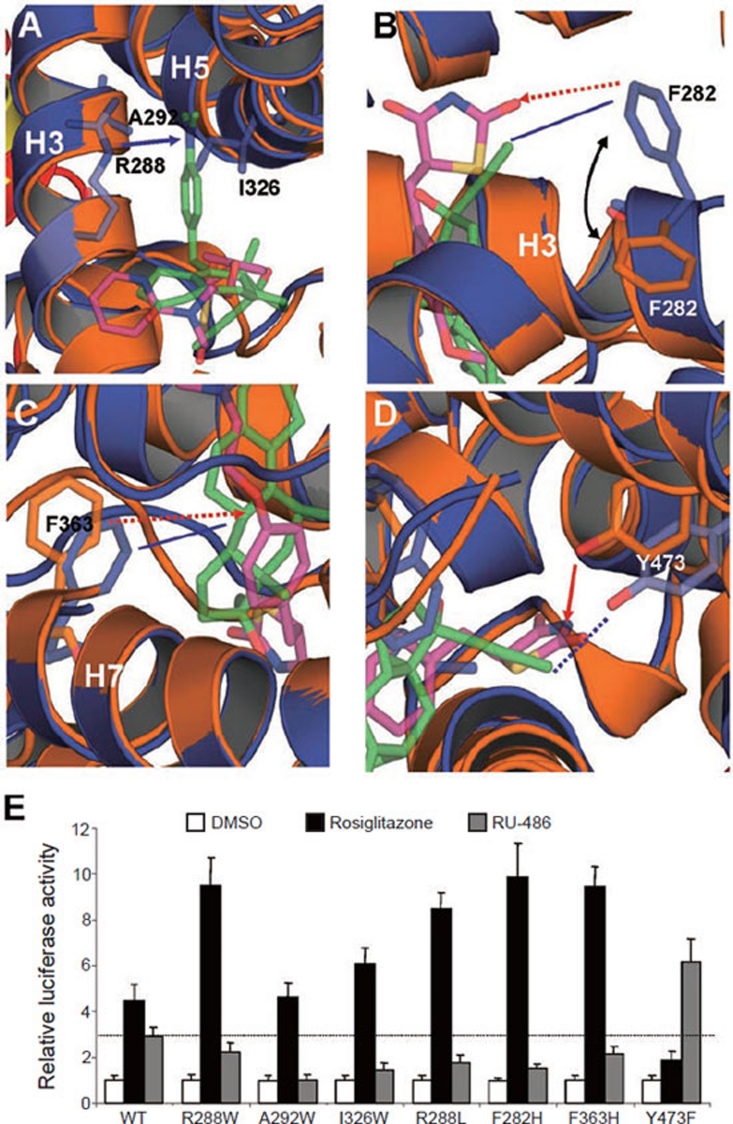
Peroxisome proliferator-activated receptor gamma (PPARγ) regulates metabolic homeostasis and is a molecular target for anti-diabetic drugs. We report here the identification of a steroid receptor ligand, RU-486, as an unexpected PPARγ agonist, thereby uncovering a novel signaling route for this steroid drug. Similar to rosiglitazone, RU-486 modulates the expression of key PPARγ target genes and promotes adipocyte differentiation, but with a lower adipogenic activity. Structural and functional studies of receptor-ligand interactions reveal the molecular basis for a unique binding mode for RU-486 in the PPARγ ligand-binding pocket with distinctive properties and epitopes, providing the molecular mechanisms for the discrimination of RU-486 from thiazolidinediones (TZDs) drugs. Our findings together indicate that steroid compounds may represent an alternative approach for designing non-TZD PPARγ ligands in the treatment of insulin resistance.


Figures

Figure 1
Figure 1
RU-486 is a high affinity PPARγ agonist. (A) Receptor-specific transactivation by RU-486. Cos7 cells were co-transfected with pG5Luc reporter together with the plasmids encoding various nuclear receptors LBDs fused with the Gal4 DNA-binding domain. After transfection, cells were treated with DMSO or 1 μM RU-486. (B) Dose responses of RU-486 in transactivating PPARγ. Cos7 cells were co-transfected with plasmids encoding full-length PPARγ and a PPRE luciferase reporter. After transfection, cells were treated with DMSO or various concentrations of RU-486 and rosiglitazone. (C) PPARγ activation by RU-486 was inhibited by PPARγ antagonist GW9662. Cos7 cells were co-transfected with plasmids encoding full-length PPARγ and a PPRE luciferase reporter. After transfection, cells were treated with RU-486, together with various concentrations of GW9662.
Figure 2
Figure 2
The transcriptional properties of PPARγ in response to RU-486 ligand. RU-486 promotes the interaction of co-activator LXXLL motifs with PPARγ. Modulation of the interaction of PPARγ LBD with various co-activator LXXLL motifs and co-repressor motifs in response to 1 μM RU-486 was shown by AlphaScreen assays. The peptide sequences are listed in experimental procedures.
Figure 3
Figure 3
RU-486 regulates PPARγ-target genes in adipogenesis and adipocytes. (A) Oil red O staining of 3T3-L1 cells after treatment with 1 μM ligands indicated for 7 days. (B) Gene expression profile during the adipocyte differentiation of 3T3-L1 cells induced by 1 μM ligands indicated.
Figure 4
Figure 4
Recognition of RU-486 by PPARγ. (A) The structures of RU-486 bound with PPARγ LBD in ribbon representation. PPARγ LBD is colored in blue and the SRC1 motif is in yellow. The bound RU-486 is shown in stick representation with carbon and oxygen atoms depicted in green and red, respectively. (B) Superposition of RU-486 (green) with rosiglitazone (purple). (C) 2Fo-Fc electron density map (1.0 σ) showing bound RU-486 to the PPARγ LBD. The bound RU-486 is shown in stick representation with carbon and oxygen atoms depicted in green and red, respectively. (D) Schematic representation of PPARγ-RU-486 interaction. The residues labeled red are unique contacts for RU-486 not observed for rosiglitazone, while the residue in blue indicates the one that lost contact with RU-486. Key hydrocarbon positions of RU-486 molecule are indicated. Hydrophobic interactions are indicated by lines and hydrogen bonds are indicated by arrows from proton donors to acceptors.
Figure 5
Figure 5
Superposition of the RU-486-bound PPARγ to rosiglitazone-bound PPARγ and RU-486-bound GR LBD. (A, B) Overlays of the PPARγ-RU486 structure (blue) with the PPARγ-rosiglitazone (gold) structure, where ligand RU-486 is in green and rosiglitazone is in purple. (C, D) Overlays of the PPARγ-RU486 structure (blue) with the GR-RU-486 structure (cyan), where ligand RU-486 is in green for PPARγ and the GR-bound RU-486 is in yellow. The dashed arrows indicate the positions of the dimethylaniline side chain of RU486.
Figure 6
Figure 6
Functional correlation of the RU-486/ PPARγ interactions. (A-D) Molecular determinants of the interaction between PPARγ with ligand RU-486. Overlays of the PPARγ-RU-486 structure (blue) with the PPARγ-rosiglitazone (gold) structure, where ligand RU-486 is in green and rosiglitazone is in purple. The hydrophobic interactions and hydrogen bonds are shown with lines and arrows, respectively. The potential hydrophobic interactions and hydrogen bonds, if the corresponding mutations are made as indicated in E, are shown in dashed lines and dashed arrows, respectively. The blue lines indicate the interactions between PPARγ and RU-486, while the gold lines indicate the interaction between PPARγ and rosiglitazone. (E) Effects of mutations of key PPARγ residues on its transcriptional activity in response to RU-486 in cell-based reporter gene assays. Cos7 cells were co-transfected with plasmids encoding full-length PPARγ or PPARγ mutants as indicated in the figure together with a PPRE luciferase reporter. The cells were treated with 1 μM RU-486 and rosiglitazone, respectively. The dashed line indicates the activation level of wild-type PPARγ by RU-486.

References

    1. Lehrke M, Lazar MA. The many faces of PPARgamma. Cell. 2005;123:993–999. - PubMed
    1. Wang YX. PPARs: diverse regulators in energy metabolism and metabolic diseases. Cell Res. 2010;20:124–137. - PMC - PubMed
    1. Yki-Jarvinen H. Thiazolidinediones. N Eng J Med. 2004;351:1106–1118. - PubMed
    1. Nissen SE, Wolski K. Effect of rosiglitazone on the risk of myocardial infarction and death from cardiovascular causes. N Eng J Med. 2007;356:2457–2471. - PubMed
    1. Waki H, Park KW, Mitro N, et al. The small molecule harmine is an antidiabetic cell-type-specific regulator of PPARgamma expression. Cell Metab. 2007;5:357–370. - PubMed

Publication types

MeSH terms

Associated data