. 2012 Jan;11(1):M111.008730.
doi: 10.1074/mcp.M111.008730. Epub 2011 Oct 10.

Tumor biomarker glycoproteins in the seminal plasma of healthy human males are endogenous ligands for DC-SIGN

Affiliations

Tumor biomarker glycoproteins in the seminal plasma of healthy human males are endogenous ligands for DC-SIGN

Gary F Clark et al. Mol Cell Proteomics. 2012 Jan.

Abstract

DC-SIGN is an immune C-type lectin that is expressed on both immature and mature dendritic cells associated with peripheral and lymphoid tissues in humans. It is a pattern recognition receptor that binds to several pathogens including HIV-1, Ebola virus, Mycobacterium tuberculosis, Candida albicans, Helicobacter pylori, and Schistosoma mansoni. Evidence is now mounting that DC-SIGN also recognizes endogenous glycoproteins, and that such interactions play a major role in maintaining immune homeostasis in humans and mice. Autoantigens (neoantigens) are produced for the first time in the human testes and other organs of the male urogenital tract under androgenic stimulus during puberty. Such antigens trigger autoimmune orchitis if the immune response is not tightly regulated within this system. Endogenous ligands for DC-SIGN could play a role in modulating such responses. Human seminal plasma glycoproteins express a high level of terminal Lewis(x) and Lewis(y) carbohydrate antigens. These epitopes react specifically with the lectin domains of DC-SIGN. However, because the expression of these sequences is necessary but not sufficient for interaction with DC-SIGN, this study was undertaken to determine if any seminal plasma glycoproteins are also endogenous ligands for DC-SIGN. Glycoproteins bearing terminal Lewis(x) and Lewis(y) sequences were initially isolated by lectin affinity chromatography. Protein sequencing established that three tumor biomarker glycoproteins (clusterin, galectin-3 binding glycoprotein, prostatic acid phosphatase) and protein C inhibitor were purified by using this affinity method. The binding of DC-SIGN to these seminal plasma glycoproteins was demonstrated in both Western blot and immunoprecipitation studies. These findings have confirmed that human seminal plasma contains endogenous glycoprotein ligands for DC-SIGN that could play a role in maintaining immune homeostasis both in the male urogenital tract and the vagina after coitus.


Figures

Fig. 1.
Fig. 1.
Analysis of DC-SIGN binding to seminal plasma glycoproteins. The seminal plasma fraction (10 μl) was subjected to SDS gel electrophoresis and stained with colloidal Coommassie blue (CCB) as shown in the panel on the left. This sample was also subjected to Western blotting with the DC-SIGN-Fc (right panel). Aliquots (5 μl) were also taken directly from fractions 1 (Fract 1) and 11 (Fract 11) obtained by chromatography on Lotus lectin-agarose (Fig. 2), and also subjected to both SDS gel electrophoresis and blotting with DC-SIGN-Fc.
Fig. 2.
Fig. 2.
Affinity chromatography of seminal plasma on Lotus lectin-agarose. Diluted seminal plasma was separated on this matrix as described in the methods. Elution with buffer containing 0.1 m fucose was initiated at the position indicated by the arrow.
Fig. 3.
Fig. 3.
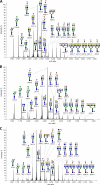
MALDI-TOF profiling of N-glycans associated with glycoprotein fractions obtained by lectin affinity chromatography of human seminal plasma. The N-glycans associated with the glycoproteins in a whole human seminal plasma fraction before it was subjected to lectin affinity chromatography as shown in Fig. 2 were determined (panel A). The N-glycans associated with the pooled unbound (panel B) and bound fractions (panel C) obtained after affinity chromatography on Lotus lectin-agarose were also defined.
Fig. 4.
Fig. 4.
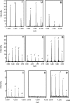
Comparison of the MALDI spectra of different classes of N-glycans associated with fractions obtained by the chromatography of whole human seminal plasma on Lotus lectin-agarose. Spectra for the N-glycans in the total (T), unbound (U) and bound seminal plasma fractions (B) obtained following lectin affinity chromatography are arranged side by side for evaluation. Signals for the high mannose type N-glycans with the formula Man5–7GlcNAc2 are indicated by notations a-c, respectively, in the top panel. In the middle panel, signals for the triantennary N-glycans bearing 2–6 fucose residues on their outer antennae are indicated by notations d-h, respectively. Tetraantennary N-glycans bearing 4–8 fucose residues in their outer antennae are indicated by the notations i-m, respectively, in the lower panel.
Fig. 5.
Fig. 5.
SDS gel electrophoretic sizing of the major seminal plasma glycoproteins bound to Lotus lectin-agarose. The bound glycoproteins eluted with 0.1 m fucose containing buffer obtained from three different samples of human seminal plasma were pooled and concentrated down to a final volume of 1 ml. A 20-μl aliquot of this fraction was subjected to SDS-gel electrophoresis and stained with colloidal Coomassie blue stain as discussed in the Methods. Individual bands were cut from this gel, and subjected to proteomic analysis as outlined in the Methods.
Fig. 6.
Fig. 6.
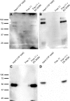
DC-SIGN binds to specific seminal plasma glycoproteins. Seminal plasma proteins eluted from the Lotus lectin affinity column (Fraction 11) were incubated with protein A-Sepharose beads either alone or in the presence of DC-SIGN-Fc. Proteins bound to beads were analyzed by Western blot with antibodies against clusterin (panel A), galectin 3-binding protein (panel B), prostatic acid phosphatase (panel C) or protein C inhibitor (panel D). Input corresponds to 1/5th of the total material present in each reaction, while the Frac. 11 lanes display the background staining contributed by nonspecific binding to the protein A-Sepharose beads.

Similar articles

Cited by

References

    1. Garcia-Vallejo J. J., van Kooyk Y. (2009) Endogenous ligands for C-type lectin receptors: the true regulators of immune homeostasis. Immunol. Rev. 230, 22–37 - PubMed
    1. Curtis B. M., Scharnowske S., Watson A. J. (1992) Sequence and expression of a membrane-associated C-type lectin that exhibits CD4-independent binding of human immunodeficiency virus envelope glycoprotein gp120. Proc. Natl. Acad. Sci. U.S.A. 89, 8356–8360 - PMC - PubMed
    1. Geijtenbeek T. B., Torensma R., van Vliet S. J., van Duijnhoven G. C., Adema G. J., van Kooyk Y., Figdor C. G. (2000) Identification of DC-SIGN, a novel dendritic cell-specific ICAM-3 receptor that supports primary immune responses. Cell 100, 575–585 - PubMed
    1. Appelmelk B. J., Van Die I., Van Vliet S. J., Vandenbroucke-Grauls C. M., Geijtenbeek T. B., Van Kooyk Y. (2003) Cutting edge: carbohydrate profiling identifies new pathogens that tnteract with dendritic cell-specific ICAM-3-grabbing nonintegrin on dendritic cells. J. Immunol. 170, 1635–1639 - PubMed
    1. Mitchell D. A., Fadden A. J., Drickamer K. (2001) A novel mechanism of carbohydrate recognition by the C-type lectins DC- SIGN and DC-SIGNR: Subunit organization and binding to multivalent ligands. J. Biol. Chem. 276, 28939–28945 - PubMed

Publication types

MeSH terms

LinkOut - more resources