. 2012 Mar;64(3):718-28.
doi: 10.1002/art.33407.

75-kd sirtuin 1 blocks tumor necrosis factor α-mediated apoptosis in human osteoarthritic chondrocytes

Affiliations

75-kd sirtuin 1 blocks tumor necrosis factor α-mediated apoptosis in human osteoarthritic chondrocytes

Hanna Oppenheimer et al. Arthritis Rheum. 2012 Mar.

Abstract

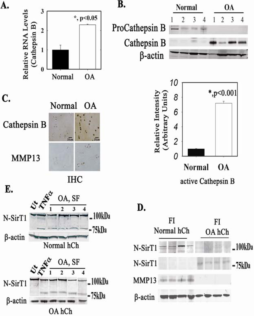
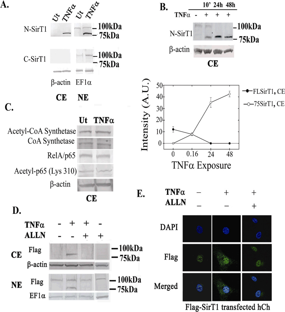
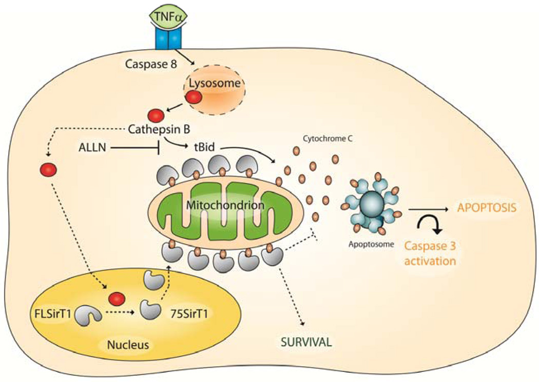
Objective: Sirtuin 1 (SirT1) has been implicated in the regulation of human cartilage homeostasis and chondrocyte survival. Exposing human osteoarthritic (OA) chondrocytes to tumor necrosis factor α (TNFα) generates a stable and enzymatically inactive 75-kd form of SirT1 (75SirT1) via cathepsin B-mediated cleavage. Because 75SirT1 is resistant to further degradation, we hypothesized that it has a distinct role in OA, and the present study was undertaken to identify this role.

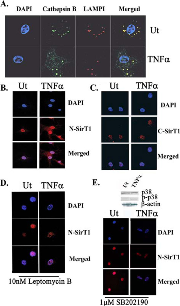
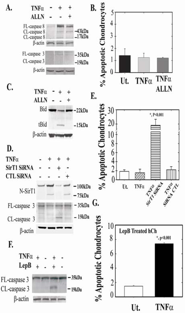
Methods: The presence of cathepsin B and 75SirT in OA and normal human chondrocytes was analyzed. Confocal imaging of SirT1 was used to monitor its subcellular trafficking following TNFα stimulation. Coimmunofluorescence staining for cathepsin B, mitochondrial cytochrome oxidase subunit IV, and lysosome-associated membrane protein 1 together with SirT1 was performed. Human chondrocytes were tested for apoptosis by fluorescence-activated cell sorter analysis and immunoblotting for caspases 3 and 8. Human chondrocyte mitochondrial extracts were obtained and analyzed for 75SirT1-cytochrome c association.

Results: Confocal imaging and immunoblot analyses following TNFα challenge of human chondrocytes demonstrated that 75SirT1 was exported to the cytoplasm and colocalized with the mitochondrial membrane. Consistent with this, immunoprecipitation and immunoblot analyses revealed that 75SirT1 is enriched in mitochondrial extracts and associates with cytochrome c following TNFα stimulation. Preventing nuclear export of 75SirT1 or reducing levels of full-length SirT1 and 75SirT1 augmented chondrocyte apoptosis in the presence of TNFα. Levels of cathepsin B and 75SirT1 were elevated in OA versus normal chondrocytes. Additional analyses showed that human chondrocytes exposed to OA-derived synovial fluid generated the 75SirT1 fragment.

Conclusion: These data suggest that 75SirT1 promotes chondrocyte survival following exposure to proinflammatory cytokines.


Conflict of interest statement

The authors declare no conflict of interest.

Figures

Figure 1
Figure 1. Osteoarthritic human chondrocytes display elevated levels of active Cathepsin B
RNA (A) and protein (B) were obtained from freshly isolated (FI) human chondrocytes (hCh) derived from normal and osteoarthritic (OA) articular cartilage (AC). n=8 separate samples per normal and OA donors. C. IHC of OA and normal AC for Cathepsin B. Note elevated Cathepsin B in OA hCh and surrounding extracellular deposits as compared to normal AC. MMP13 IHC served as a marker for osteoarthritic AC in each equivalent sample. n=4 with 2 replicates per sample. D. Immunoblots of FI hCh from normal and OA cartilage were run with N-SirT1 antibody, n=8 separate samples per normal and OA donors. E. Normal (upper panel) and OA hCh (lower panel) were stimulated with TNFα and synovial fluid (SF, 10v% in BIO-MPM serum-free medium) derived from OA joints. Following 24h incubation, protein extracts were obtained and immunoblotted. Results in E, were repeated with three different origins of normal and OA hCh samples and four different SF samples from OA patients. "Ut" denotes untreated hCh and "TNFα" denotes TNFα treated (50 ng/mL, 24h) hCh. Immunoblots indicate 75 and 100 kDa protein ladder, on the right hand side.
Figure 2
Figure 2. N-terminally intact SirT1 is exported to the cytoplasm in a MAPK p38 and CRM1-dependant fashion, following TNFα-treatment
A. Confocal images of LAMPI (red fluorescence), active Cathepsin B (Cathepsin B; green fluorescence) and DAPI (blue fluorescence) TNFα treated (TNFα) and untreated (Ut) samples. B. Confocal images of hCh stained with an N-SirT1 antibody (red fluorescence). The nucleus was stained with DAPI, which appears in blue fluorescence. C. Confocal images of C- SirT1 (red fluorescence) and DAPI D. Human chondrocytes were treated with Leptomycin B (10nM LepB), a CRM1 (Exportin-1) inhibitor, for 3h prior TNFα treatment. The cells were then stained with an N-SirT1 antibody (red fluorescence) and DAPI (blue fluorescence). E. Immunoblots of MAPK p38 and phospho-p38 (upper panel) in untreated and TNFα-treated hCh. Lower panel exhibit hCh treated with SB202190 (1µg/mL), a MAPK p38 inhibitor, with and without TNFα stimulation. The samples were then stained with N-SirT1 antibody (red fluorescence) and DAPI. Results were repeated in duplicate samples of three separate OA hCh origins (n=3).
Figure 3
Figure 3. 75SirT1 fragment is exported to the cytoplasm upon TNFα stimulation
A. Isolated CE and NE were immunoblotted for N-SirT1 (upper panel) and C-SirT1 antibody (lower panel). B. OA hCh are treated with 50 ng/mL TNFα for 10 min, 24h and 48h incubation, as indicated above the blot. CE were isolated and immunoblotted with N-SirT1 antibody. Lower panel indicated band intensity in arbitrary units (AU) of 75SirT1 (white circles) and FLSirT1 (black circles) as a function of TNFα time exposure. C. CE protein extracts were obtained and immunoblotted for cytoplasmic protein targets of SirT1 deacetylation. E. OA hCh were transfected with a SirT1 expression plasmid possessing a Flag tag on its N-terminal end (Flag-SirT1) and subsequently treated with TNFα and/or ALLN. Thereafter CE and NE were run for Flag antibody. F. Human OA chondrocytes were treated as indicated in E, stained for Flag antibody (green fluorescence) and DAPI (blue fluorescence) and inspected via confocal microscope. "Ut" denotes untreated hCh and "TNFα" denotes TNFα treated (50ng/mL, 24h) hCh. Immunoblots indicate 75 and 100 kDa protein ladder on the right hand side. Results were repeated in duplicate samples of three separate OA hCh origins (n=3).
Figure 4
Figure 4. 75SirT1 colocalizes with the mitochondrial membrane and Cytochrome C following TNFα treatment
OA hCh were treated with TNFα (TNFα) or untreated (Ut), as indicated. A. Confocal images stained with Cytochrome Oxidase Subunit IV (Cox IV; green fluorescence) and N-SirT1 (red fluorescence) antibodies display enhanced colocalization of cytoplamic 75SirT1 on the mitochondrial membrane, following TNFα stimuli. B. Mitochondrial protein extracts (ME) were immunoblotted for N-SirT1 (upper panel), CoxIV and Cytochrome C. C. MEs of TNFα-treated (TNFα) or untreated (Ut) OA hCh were obtained, immunoprecipitated with endogenous N-SirT1 and subsequently immunoblotted for both N-SirT1 and Cytochrome C antibodies. D. Human OA chondrocytes were transfected with Flag-SirT1 or pcDNA and treated with TNFα. Whole cell extract (WCE) inputs confirmed elevated SirT1 levels as compared to pcDNA derived extracts (upper panel). The lower panel of D displays Flag immunoprecipitants (IP:Flag) from TNFα-treated WCE, which were subsequently immunoblotted for Flag and Cytochrome C. Immunoblots indicate 75 and 100 kDa protein ladder on the right hand side. Results were repeated in duplicate samples of three separate OA hCh origins (n=3).
Figure 5
Figure 5. 75SirT1 promotes human chondrocyte survival
OA Human chondrocytes were treated as indicated above the immunoblots. A. Following treatment, whole cell extract (WCE) were immunoblotted for cleaved (CL) and full-length (FL) caspase 8 and 3. B. FACS analyses for apoptotic cell population was carried out using double staining for Annexin V and propidium iodide, for OA hCh treated as indicated in A. C. hCh were treated as indicated above the blot and run for Bid and tBid. D. Human chondrocytes were treated with SirT1 SiRNA, control (CTL) SiRNA, with or without TNFα (as indicated above the immunoblot). WCE were obtained and immunoblotted with N-SirT1 antibody, CL and FL caspase 3. E. FACS analyses for OA hCh in C. F. Human chondrocytes were treated with the CRM1 inhibitor, Leptomycin B (LepB; 10nM) and TNFα, as indicated above the immunoblot. WCE were immunoblotted for FL and CL caspase 3. G. FACS analyses for hCh treated as in E. FACS analyses were repeated twice in three different samples. Results determined significance according to t-test at p<0.001 confidence Immunoblots indicate 75 and 100 kDa protein ladder on the right hand side. Results were repeated in duplicate samples of three separate OA hCh origins (n=3).
Figure 6
Figure 6. Predicted role of 75SirT1 in chondrocyte survival
Following TNFα stimulation caspase 8 dependant lysosomal permeability occurs. Lysosomal permeability exerts augmented levels of Cathepsin B in the cell cytoplasm and nucleus. Active Cathepsin B cleaves nuclear FLSirT1 to generate an inactive stable 75SirT1 fragment, which is exported via CRM1 to the cytoplasm (broken arrows). While in the Cytoplasm 75SirT1 interacts with Cytochrome C on the mitochondrial membrane to block downstream apoptososme assembly (broken arrows). Solid black arrows indicate the TNFα-Cathepsin B pathways through Bid activation. ALLN will block both Bid cleavage and 75SirT1 generation. The model may be relevant in-vivo for articular chondrocytes prone to develop OA (see SD-6).

References

    1. Iannone F, Lapadula G. The pathophysiology of osteoarthritis. Aging Clin Exp Res. 2003;15:364–372. - PubMed
    1. Goldring SR, Goldring MB. Clinical aspects, pathology and pathophysiology of osteoarthritis. J Musculoskelet Neuronal Interact. 2006;6:376–378. - PubMed
    1. Petersson IF, Jacobsson LT. Osteoarthritis of the peripheral joints. Best Pract Res Clin Rheumatol. 2002;16:741–760. - PubMed
    1. Loeser RF. Age-related changes in the musculoskeletal system and the development of osteoarthritis. Clin Geriatr Med. 2010;26(3):371–386. - PMC - PubMed
    1. Fernandes JC, Martel-Pelletier J, Pelletier JP. The role of cytokines in osteoarthritis pathophysiology. Biorheology. 2002;39:237–246. - PubMed

Publication types

MeSH terms