. 2011 Nov 25;286(47):40717-24.
doi: 10.1074/jbc.M111.289975. Epub 2011 Oct 10.

Activity of 3-ketosteroid 9α-hydroxylase (KshAB) indicates cholesterol side chain and ring degradation occur simultaneously in Mycobacterium tuberculosis

Affiliations

Activity of 3-ketosteroid 9α-hydroxylase (KshAB) indicates cholesterol side chain and ring degradation occur simultaneously in Mycobacterium tuberculosis

Jenna K Capyk et al. J Biol Chem. .

Abstract

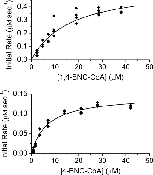
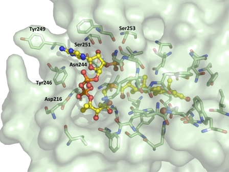
Mycobacterium tuberculosis (Mtb), a significant global pathogen, contains a cholesterol catabolic pathway. Although the precise role of cholesterol catabolism in Mtb remains unclear, the Rieske monooxygenase in this pathway, 3-ketosteroid 9α-hydroxylase (KshAB), has been identified as a virulence factor. To investigate the physiological substrate of KshAB, a rhodococcal acyl-CoA synthetase was used to produce the coenzyme A thioesters of two cholesterol derivatives: 3-oxo-23,24-bisnorchol-4-en-22-oic acid (forming 4-BNC-CoA) and 3-oxo-23,24-bisnorchola-1,4-dien-22-oic acid (forming 1,4-BNC-CoA). The apparent specificity constant (k(cat)/K(m)) of KshAB for the CoA thioester substrates was 20-30 times that for the corresponding 17-keto compounds previously proposed as physiological substrates. The apparent K(m)(O(2)) was 90 ± 10 μM in the presence of 1,4-BNC-CoA, consistent with the value for two other cholesterol catabolic oxygenases. The Δ(1) ketosteroid dehydrogenase KstD acted with KshAB to cleave steroid ring B with a specific activity eight times greater for a CoA thioester than the corresponding ketone. Finally, modeling 1,4-BNC-CoA into the KshA crystal structure suggested that the CoA moiety binds in a pocket at the mouth of the active site channel and could contribute to substrate specificity. These results indicate that the physiological substrates of KshAB are CoA thioester intermediates of cholesterol side chain degradation and that side chain and ring degradation occur concurrently in Mtb. This finding has implications for steroid metabolites potentially released by the pathogen during infection and for the design of inhibitors for cholesterol-degrading enzymes. The methodologies and rhodococcal enzymes used to generate thioesters will facilitate the further study of cholesterol catabolism.


Figures

FIGURE 1.
FIGURE 1.
Schematic of the proposed cholesterol degradation pathway of Mtb. Key carbons of cholesterol are numbered using the steroid numbering convention. The processes of cholesterol side chain and partial ring degradation are shown as parallel pathways. Enzymes known to catalyze specific reactions in Mtb are indicated. Non-enzymatic reaction is shown as a dashed arrow. Compounds 1, 2, and 3 represent 4-AD, ADD, and 3-hydroxy-9,10-seconandrost-1,3,5(10)-trien-9,17-dione (3-HSA) (R = ketone); 4-BNC, 1,4-BNC, and 3-HSBNC, (R = isopropionate); 4-BNC-CoA, 1,4-BNC-CoA, and 3-HSBNC-CoA, (R = isopropionyl-CoA).
FIGURE 2.
FIGURE 2.
Compounds investigated as KshAB substrates. Shown are 4-AD (A), ADD (B), 4-BNC (C), 1,4-BNC (D), and 1,4-BNC-CoA (E). Not shown is 4-BNC-CoA, which differs from E by a 1–2 saturated bond on ring A.
FIGURE 3.
FIGURE 3.
Transformation of 4-BNC to 4-BNC-CoA by CasI. HPLC chromatograms are shown of 50 μl of a CasI reaction (2 μm, dashed line) and no-enzyme control (solid line) containing 0.9 mm 4-BNC, 1.1 mm CoASH, 5 mm MgCl2, and 2.5 mm ATP after 7 h. Peaks were resolved using a linear gradient of 0 to 90% methanol in 0.1 m ammonium acetate, pH 4.5, over 20 min. Substrate peaks are numbered: 1, ATP; 3, CoASH; 5, 4-BNC. Product peaks are numbered: 2, AMP; 4, 4-BNC-CoA.
FIGURE 4.
FIGURE 4.
Steady-state kinetic analyses of KshAB with CoA thioesters. Shown is the dependence of the initial velocity of O2 consumption on 1,4-BNC-CoA (A) and 4-BNC-CoA (B) concentrations in air-saturated buffer with fitted parameters Km = 17 ± 3 and 6.8 ± 0.1 μm, and Vmax = 0.56 ± 0.04 and 0.146 ± 0.007 μm s−1, respectively. The best fit of the Michaelis-Menten equation to the data using the least squares dynamic weighting options of LEONORA is represented as a solid line.
FIGURE 5.
FIGURE 5.
Docking of 1,4-BNC-CoA in active site of KshA. 1,4-BNC-CoA is shown as a ball and stick representation. KshA (PDB ID 2ZYL (15)) is shown as a green, semitransparent surface. KshA amino acids within 4 Å of the CoA group of 1,4-BNC-CoA are shown as sticks and labeled. Carbon atoms of amino acid residues and the substrate are colored green and yellow, respectively. Oxygen, phosphorus, and nitrogen atoms are shown in red, orange, and blue, respectively. The catalytic iron is shown as a rust-colored sphere. Residues mentioned in the text are labeled.

References

    1. WHO (2010) Global Tuberculosis Control: WHO Report 2010, pp. 2–42, World Health Organization, Geneva, Switzerland
    1. Russell D. G. (2007) Nat. Rev. Microbiol 5, 39–47 - PubMed
    1. Russell D. G., VanderVen B. C., Lee W., Abramovitch R. B., Kim M. J., Homolka S., Niemann S., Rohde K. H. (2010) Cell Host Microbe 8, 68–76 - PMC - PubMed
    1. Schnappinger D., Ehrt S., Voskuil M. I., Liu Y., Mangan J. A., Monahan I. M., Dolganov G., Efron B., Butcher P. D., Nathan C., Schoolnik G. K. (2003) J. Exp. Med. 198, 693–704 - PMC - PubMed
    1. Homolka S., Niemann S., Russell D. G., Rohde K. H. (2010) PLoS Pathog. 6, e1000988 - PMC - PubMed

Publication types

MeSH terms