

Identification and enumeration of circulating tumor cells in the cerebrospinal fluid of breast cancer patients with central nervous system metastases
- PMID: 21987585
- PMCID: PMC3248154
- DOI: 10.18632/oncotarget.336
Identification and enumeration of circulating tumor cells in the cerebrospinal fluid of breast cancer patients with central nervous system metastases
Abstract
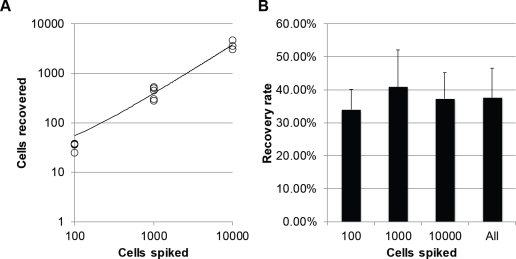
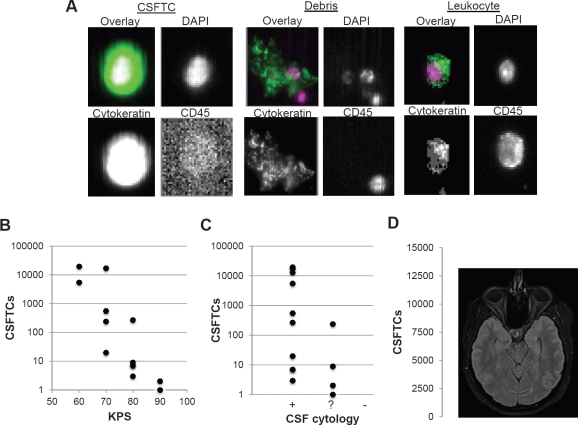
The number of circulating tumor cells (CTCs) in the peripheral blood of metastatic breast cancer patients is now an established prognostic marker. While the central nervous system is a common site of metastasis in breast cancer, the standard marker for disease progression in this setting is cerebrospinal fluid (CSF) cytology. However, the significance of CSF cytology is unclear, requires large sample size, is insensitive and subjective, and sometimes yields equivocal results. Here, we report the detection of breast cancer cells in CSF using molecular markers by adapting the CellSearch system (Veridex). We used this platform to isolate and enumerate breast cancer cells in CSF of breast cancer patients with central nervous system (CNS) metastases. The number of CSF tumor cells correlated with tumor response to chemotherapy and were dynamically associated with disease burden. This CSF tumor cell detection method provides a semi-automated molecular analysis that vastly improves the sensitivity, reliability, objectivity, and accuracy of detecting CSF tumor cells compared to CSF cytology. CSF tumor cells may serve as a marker of disease progression and early-stage brain metastasis in breast cancer and potentiate further molecular analysis to elucidate the biology and significance of tumor cells in the CSF.
Figures


Comment in
-
Circulating tumor cells in the cerebrospinal fluid: "tapping" into diagnostic and predictive potential.Oncotarget. 2011 Nov;2(11):822. doi: 10.18632/oncotarget.349. Oncotarget. 2011. PMID: 22064881 Free PMC article. No abstract available.
References
-
- Cristofanilli M, Budd GT, Ellis MJ, Stopeck A, Matera J, Miller MC, Reuben JM, Doyle GV, Allard WJ, Terstappen LW, Hayes DF. Circulating tumor cells, disease progression, and survival in metastatic breast cancer. New Engand Journal of Medicine. 2004;351:781–91. - PubMed
-
- Paterlini-Brechot P, Benali NL. Circulating tumor cells (CTC) detection: clinical impact and future directions. Cancer Letters. 2007;253:180–204. - PubMed
-
- Glantz MJ, Van Horn A, Fisher R, Chamberlain MC. Route of intracerebrospinal fluid chemotherapy administration and efficacy of therapy in neoplastic meningitis. Cancer. 2010;116:1947–52. - PubMed
-
- Heitz F, Rochon J, Harter P, Lueck HJ, Fisseler-Eckhoff A, Barinoff J, Traut A, Lorenz-Salehi F, du Bois A. Cerebral metastases in metastatic breast cancer: disease-specific risk factors and survival. Annals of Oncology. 2011;22:1571–81. - PubMed
-
- Mori T, Sugita K, Kimura K, Fuke T, Miura T, Kiyokawa N, Fujimoto J. Isolated central nervous system (CNS) relapse in a case of childhood systemic anaplastic large cell lymphoma without initial CNS involvement. Journal of Pediatric Hematology/Oncology. 2003;25:975–7. - PubMed
MeSH terms
Substances
LinkOut - more resources
Full Text Sources
Other Literature Sources
Medical
