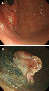
Do you have what it takes for challenging endoscopic submucosal dissection cases?
- PMID: 21987603
- PMCID: PMC3180013
- DOI: 10.3748/wjg.v17.i31.3580
Do you have what it takes for challenging endoscopic submucosal dissection cases?
Abstract
Endoscopic submucosal dissection (ESD) is a widely accepted treatment for early gastric cancer (EGC), especially in Korea and Japan. The criteria for the therapeutic use of ESD for EGC have been expanded recently. However, attention should be drawn to the technical feasibility of the ESD treatment which depends on a lesion's location, size or fibrosis level, or operator's experience. In the case of a lesion with a high level of difficulty, a more experienced operator is required. Thus, the treatment for a lesion with a high level of difficulty should be performed according to the degree of the operator's experience. In this paper, the authors describe the ESD procedure for lesions with a high level of difficulty.
Keywords: Anatomical location; Endoscopic submucosal dissection; Technical feasibility.
Figures


References
-
- Cho JY, Cho WY. The current status of endoscopic submucosal dissection. Korean J Gastrointest Endosc. 2008;37:317–320 (Korean).
-
- Gotoda T, Yamamoto H, Soetikno RM. Endoscopic submucosal dissection of early gastric cancer. J Gastroenterol. 2006;41:929–942. - PubMed
-
- Kim JJ, Lee JH, Jung HY, Lee GH, Cho JY, Ryu CB, Chun HJ, Park JJ, Lee WS, Kim HS, et al. EMR for early gastric cancer in Korea: a multicenter retrospective study. Gastrointest Endosc. 2007;66:693–700. - PubMed
-
- Goto O, Fujishiro M, Kodashima S, Ono S, Omata M. Is it possible to predict the procedural time of endoscopic submucosal dissection for early gastric cancer? J Gastroenterol Hepatol. 2009;24:379–383. - PubMed
-
- Imagawa A, Okada H, Kawahara Y, Takenaka R, Kato J, Kawamoto H, Fujiki S, Takata R, Yoshino T, Shiratori Y. Endoscopic submucosal dissection for early gastric cancer: results and degrees of technical difficulty as well as success. Endoscopy. 2006;38:987–990. - PubMed
MeSH terms
LinkOut - more resources
Full Text Sources
Medical
Miscellaneous

