

Outcome after endoscopic submucosal dissection for early gastric cancer in Korea
- PMID: 21987605
- PMCID: PMC3180015
- DOI: 10.3748/wjg.v17.i31.3591
Outcome after endoscopic submucosal dissection for early gastric cancer in Korea
Abstract
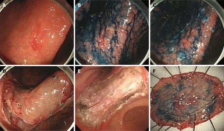
Endoscopic treatment, such as endoscopic mucosal resection (EMR) and endoscopic submucosal dissection (ESD), has been established as one of the treatment options for selected cases with early gastric cancer (EGC). Most studies on this topic have been carried out by researchers in Japan. Recently, the experience in EMR/ESD for EGC outside Japan is increasingly reported. In Korea, gastric cancer is the most common malignant disease, and the second leading cause of cancer death. Currently, EMR for EGC is widely performed in many centers in Korea. Early results with a short-term follow-up period are very promising in Korea. The complete resection rate of EMR was 37.8%-94.3%, and that of ESD was 77.4%-93.1%. In this review, we will provide an overview of the outcomes of endoscopic treatments in Korea.
Keywords: Early gastric cancer; Endoscopic mucosal resection; Endoscopic submucosal dissection; Outcome.
Figures
Similar articles
-
Endoscopic submucosal dissection for early gastric cancer: quo vadis?World J Gastroenterol. 2011 Jun 7;17(21):2623-5. doi: 10.3748/wjg.v17.i21.2623. World J Gastroenterol. 2011. PMID: 21677830 Free PMC article.
-
Endoscopic resection for early gastric cancer: current status in Korea.Dig Endosc. 2012 May;24 Suppl 1:159-65. doi: 10.1111/j.1443-1661.2012.01275.x. Dig Endosc. 2012. PMID: 22533774
-
Worldwide experiences of endoscopic submucosal dissection: not just Eastern acrobatics.World J Gastroenterol. 2011 Jun 7;17(21):2611-7. doi: 10.3748/wjg.v17.i21.2611. World J Gastroenterol. 2011. PMID: 21677828 Free PMC article.
-
Endoscopic mucosal resection of early gastric cancer: Experiences in Korea.World J Gastroenterol. 2007 Jul 21;13(27):3657-61. doi: 10.3748/wjg.v13.i27.3657. World J Gastroenterol. 2007. PMID: 17659722 Free PMC article. Review.
-
Endoscopic submucosal dissection of early gastric cancer.Digestion. 2008;77 Suppl 1:23-8. doi: 10.1159/000111484. Epub 2008 Jan 18. Digestion. 2008. PMID: 18204258 Review.
Cited by
-
Impact of tumor location on clinical outcomes of gastric endoscopic submucosal dissection.World J Gastroenterol. 2014 Jul 14;20(26):8631-7. doi: 10.3748/wjg.v20.i26.8631. World J Gastroenterol. 2014. PMID: 25024619 Free PMC article.
-
Long-Term Follow-Up After Non-Curative Endoscopic Submucosal Dissection for Early Gastrointestinal Cancer-A Retrospective Multicenter Analysis.J Clin Med. 2024 Nov 2;13(21):6594. doi: 10.3390/jcm13216594. J Clin Med. 2024. PMID: 39518733 Free PMC article.
-
Is Radical Surgery Necessary for All Patients Diagnosed as Having Non-Curative Endoscopic Submucosal Dissection?Clin Endosc. 2019 Jan;52(1):21-29. doi: 10.5946/ce.2019.014. Epub 2019 Jan 30. Clin Endosc. 2019. PMID: 30727716 Free PMC article.
-
Endoscopic ultrasonography for staging of T1a and T1b esophageal squamous cell carcinoma.World J Gastroenterol. 2014 Feb 7;20(5):1340-7. doi: 10.3748/wjg.v20.i5.1340. World J Gastroenterol. 2014. PMID: 24574809 Free PMC article.
-
Endoscopic Submucosal Dissection in the Upper Gastrointestinal Tract and the Need for Rescue Surgery-A Multicenter Analysis.J Clin Med. 2023 Nov 6;12(21):6940. doi: 10.3390/jcm12216940. J Clin Med. 2023. PMID: 37959405 Free PMC article.
References
-
- Choi IJ. [Gastric cancer screening and diagnosis] Korean J Gastroenterol. 2009;54:67–76. - PubMed
-
- Cho J, Guallar E, Hsu YJ, Shin DW, Lee WC. A comparison of cancer screening practices in cancer survivors and in the general population: the Korean national health and nutrition examination survey (KNHANES) 2001-2007. Cancer Causes Control. 2010;21:2203–2212. - PubMed
-
- Cho JY, Cho WY. Toward the global standardization of endoscopic submucosal dissection proposal for 10 years from now - present and future view of Korea. Dig Endosc. 2009;21 Suppl 1:S2–S3. - PubMed
-
- Soetikno R, Kaltenbach T, Yeh R, Gotoda T. Endoscopic mucosal resection for early cancers of the upper gastrointestinal tract. J Clin Oncol. 2005;23:4490–4498. - PubMed
-
- Hirooka Y, Naitoh Y, Goto H, Ito A, Hayakawa S, Watanabe Y, Ishiguro Y, Kojima S, Hashimoto S, Hayakawa T. Contrast-enhanced endoscopic ultrasonography in gallbladder diseases. Gastrointest Endosc. 1998;48:406–410. - PubMed
MeSH terms
LinkOut - more resources
Full Text Sources
Medical
Miscellaneous
