. 2011 Oct 17;195(2):231-43.
doi: 10.1083/jcb.201107086. Epub 2011 Oct 10.

The Protein Phosphatase 2A regulatory subunit Twins stabilizes Plk4 to induce centriole amplification

Affiliations

The Protein Phosphatase 2A regulatory subunit Twins stabilizes Plk4 to induce centriole amplification

Christopher W Brownlee et al. J Cell Biol. .

Abstract

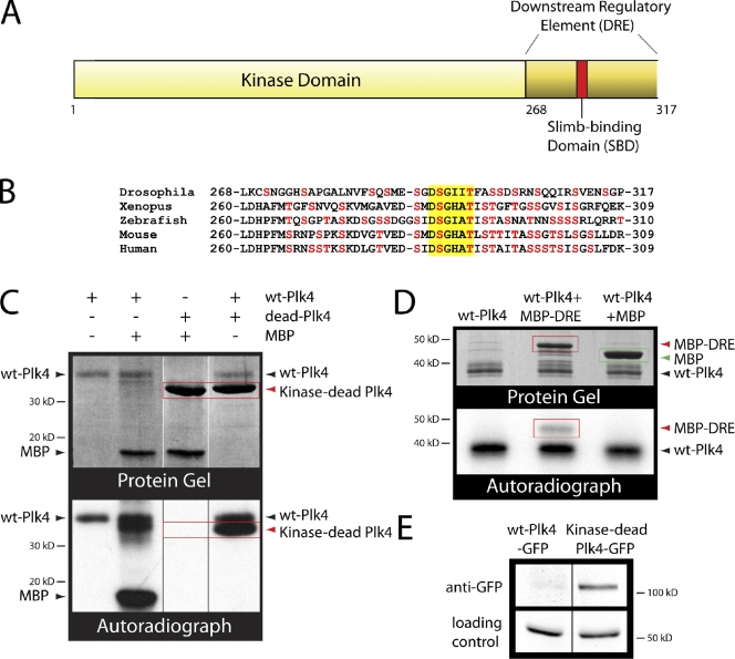
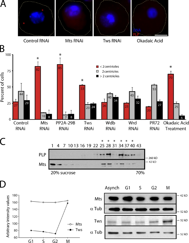
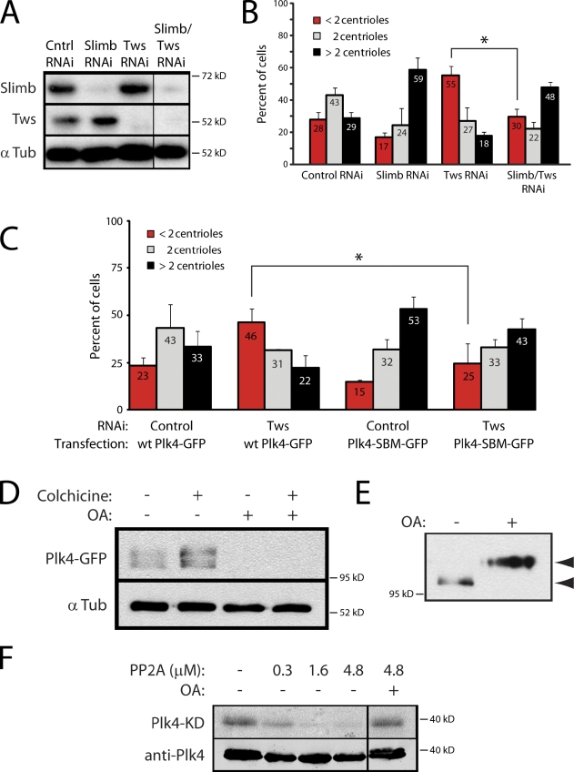
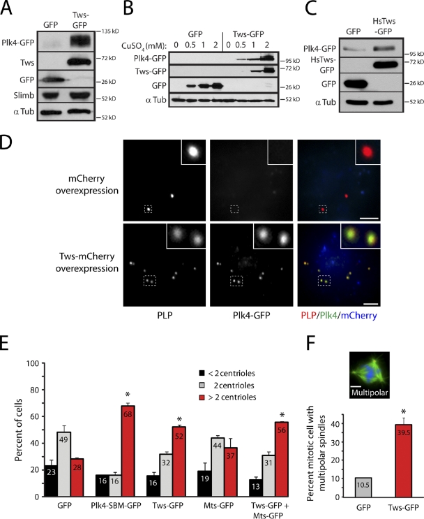
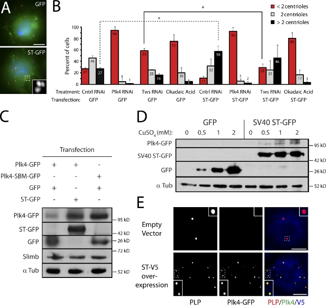
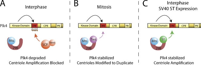
Centriole duplication is a tightly regulated process that must occur only once per cell cycle; otherwise, supernumerary centrioles can induce aneuploidy and tumorigenesis. Plk4 (Polo-like kinase 4) activity initiates centriole duplication and is regulated by ubiquitin-mediated proteolysis. Throughout interphase, Plk4 autophosphorylation triggers its degradation, thus preventing centriole amplification. However, Plk4 activity is required during mitosis for proper centriole duplication, but the mechanism stabilizing mitotic Plk4 is unknown. In this paper, we show that PP2A (Protein Phosphatase 2A(Twins)) counteracts Plk4 autophosphorylation, thus stabilizing Plk4 and promoting centriole duplication. Like Plk4, the protein level of PP2A's regulatory subunit, Twins (Tws), peaks during mitosis and is required for centriole duplication. However, untimely Tws expression stabilizes Plk4 inappropriately, inducing centriole amplification. Paradoxically, expression of tumor-promoting simian virus 40 small tumor antigen (ST), a reported PP2A inhibitor, promotes centrosome amplification by an unknown mechanism. We demonstrate that ST actually mimics Tws function in stabilizing Plk4 and inducing centriole amplification.


Figures

Figure 1.
Figure 1.
Drosophila Plk4 autophosphorylation promotes its own degradation. (A) Linear map of Drosophila Plk4 amino-terminus encoding the KD and the DRE. The red bar indicates the position of the conserved SBD (DSGXXT). (B) Lineup of the 50 amino acid DRE encoded by Plk4 family members. Serine and threonine residues are shown in red. Yellow box highlights the SBD. (C) Purified recombinant Plk4 kinase domain + DRE (wt-Plk4) but not kinase-dead (D156N point mutation of wt) Plk4 autophosphorylates in vitro. Coomassie-stained gel (top) and corresponding autoradiograph (bottom) are shown. (lanes 1 and 2) Plk4 phosphorylates itself and purified bovine myelin basic protein (MBP). (lane 3) Kinase-dead Plk4 lacks kinase activity. (lane 4) wt-Plk4 phosphorylates kinase-dead Plk4 in trans. (D) wt-Plk4 phosphorylates purified DRE fused to maltose-binding protein (MBP-DRE) in trans but does not phosphorylate purified maltose-binding protein. (E) A kinase-dead mutation in Plk4 inhibits its degradation in S2 cells. Anti-GFP immunoblot of S2 cell lysates shows that full-length kinase-dead Plk4-GFP is more stable than wt-Plk4-GFP, which is degraded and nearly undetectable. Inducible Plk4 constructs were cotransfected into S2 cells with Nlp-GFP (used as a loading control and driven by its endogenous promoter). Black/white lines indicate that intervening lanes have been spliced out.
Figure 2.
Figure 2.
PP2ATws is required for centriole duplication. (A) 5-d RNAi-treated or 24-h OA-treated S2 cells were immunostained for PLP to mark centrioles and Hoechst stained to label DNA. Cell borders are traced with dashed lines. Bar, 5 µm. (B) PP2A inhibition by Mts, 29B, or Tws RNAi leads to centriole loss. After RNAi treatment, the number of PLP-immunostained centrioles per cell was measured. Graph shows the percentage of cells with the indicated number of centrioles per cell. Each number in a bar is the percent mean for two experiments (n = 500 cells/treatment). Asterisks mark significant differences (relative to control) for comparisons mentioned in the text (P < 0.02). Error bars indicate SD. (C) Mts cosediments with centrioles purified from mitotic S2 cells on a 20–70% sucrose gradient. Fractions (numbered) were immunoblotted for the indicated proteins. Asterisks mark the major centriole-containing fractions. (D) Tws protein is maximal during mitosis. (left) Graph of normalized endogenous Mts and Tws levels in asynchronous cells (Asynch) or after a 24-h drug-induced cell cycle arrest. Plotted values were determined from the anti-Mts and Tws immunoblots (right) shown. The graph and blots are representative examples of three independent experiments, all with similar results. α-Tubulin (α Tub) was used as a loading control. Wdb, Widerborst; Wrd, Well rounded.
Figure 3.
Figure 3.
PP2ATws stabilizes Plk4 to promote centriole duplication. (A) Immunoblots of 6-d RNAi-treated S2 cells demonstrating knockdown of target proteins. α Tub, α-tubulin; Cntrl, control. (B) Loss of centrioles by Tws RNAi is rescued by codepletion of Slimb. Each mean percentage of cells (numbers) is derived from two experiments (n = 598 cells/treatment). Asterisk indicates significant difference (P < 0.02) between compared treatments mentioned in the text. (C) Low expression of nondegradable Plk4-SBM-GFP also rescues the centriole loss by Tws-RNAi. Each mean percentage (numbers) is derived from three experiments (n = 900 cells/treatment). *, P < 0.04. (D) PP2A is required to stabilize Plk4. S2 cells overexpressing Plk4-GFP were treated with colchicine or OA for 24 h as indicated, and lysates were probed for GFP. (E) PP2A dephosphorylates Plk4-SBM-GFP in cells. S2 cells expressing Plk4-SBM-GFP were treated with OA for 24 h, and their lysates were immunoblotted for GFP. Note the clear shift in mobility of Plk4-SBM after OA treatment (arrowheads), consistent with Plk4-SBM being hyperphosphorylated after PP2A inhibition. (F) PP2A dephosphorylates Plk4 in vitro. Human PP2A dephosphorylates autophosphorylated Plk4-KD + DRE-(His)6 protein (Plk4-KD) but is inhibited by OA. (top) Autoradiogram; (bottom) Plk4 immunoblot. Black lines indicate that intervening lanes have been spliced out. Error bars indicate SD.
Figure 4.
Figure 4.
Ectopic Tws expression is sufficient to stabilize Plk4 and promote centriole amplification. (A) Immunoblots of S2 cell lysates showing that Tws-GFP overexpression stabilizes Plk4-GFP without affecting Slimb levels. S2 cells were cotransfected with inducible Plk4-GFP and inducible GFP (first lane) or Tws-GFP (second lane) expression constructs. After a 24-h recovery period, gene expression was induced with 1 mM CuSO4 for 20 h. Cell lysates were then prepared and immunoblotted for GFP, endogenous Slimb, and α-tubulin (α Tub; loading control). (B) The extent of Plk4-GFP stabilization is dose dependent on Tws-GFP. S2 cells were cotransfected with Plk4-GFP (driven by the weak, constitutive Drosophila SAS-6 promoter) and either GFP (negative control) or Tws-GFP, each controlled by the copper-inducible metallothionein promoter. Cells were incubated with 0, 0.5, 1, or 2 mM CuSO4 for 24 h, and cell lysates were probed by GFP immunoblotting. (C) Immunoblot of S2 cell lysates showing that Plk4-GFP is also stabilized by human Tws (HsTws; PR55-α) overexpression. (D) Tws-mCherry expression drives abnormal accumulation of Plk4-GFP on centrioles (anti-D-PLP) in interphase S2 cells. Insets show centrioles (dashed boxes) at higher magnification. Bars, 5 µm. (E) Tws-GFP overexpression promotes centriole amplification. Graph shows the percentage of transgene-expressing cells containing the indicated number of centrioles; means (numbers) derived from three experiments (n = 600 cells/treatment). *, P < 0.003 (treated conditions compared with GFP control). (F) Tws-GFP overexpression increases the frequency of multipolar spindles. S2 cells expressing GFP or Tws-GFP were immunostained for centrioles (anti-PLP, red) and α-tubulin (green). DNA (blue) is Hoechst stained. Bar, 2.5 µm. Graph shows mean percentages (numbers) of mitotic cells with multipolar spindles (two experiments; n = 76 cells/treatment). *, P < 0.01. Error bars indicate SD.
Figure 5.
Figure 5.
Tumor-promoting SV40 ST mimics Tws function to stabilize Plk4 and amplify centrioles. (A) S2 cells overexpressing GFP or ST-GFP (green) immunostained for PLP (red) to mark centrioles. DNA (blue) is Hoechst stained. (B) ST-GFP overexpression increases centriole numbers but not in the presence of OA. ST-GFP expression also rescues centriole loss caused by depletion of Tws but not Plk4. Centrioles were counted after 6-d RNAi or 24-h OA treatment in cells transfected with inducible GFP or ST-GFP (expression was induced during the last 2 d). Mean values (numbers) of two experiments are shown (n = 600 cells/treatment). Asterisks indicate significant differences (P < 0.05) between compared treatments mentioned in the text. Error bars indicate SD. Ctnrl, control. (C and D) Immunoblots show that ST-GFP overexpression stabilizes Plk4-GFP in S2 cells to levels similar to Plk4-SBM-GFP, without affecting endogenous Slimb levels (C) and in a dose-dependent manner (D; treatment protocol similar to that described in Fig. 4 B). α Tub, α-tubulin. (E) ST-V5 expression drives accumulation of Plk4-GFP on centrioles (anti-PLP) in interphase S2 cells. Insets show centrioles (dashed boxes) at higher magnification. Bars, 5 µm.
Figure 6.
Figure 6.
Model depicting the regulation of Plk4 activity by a counteracting autophosphorylation/dephosphorylation mechanism. (A) Plk4 is expressed throughout interphase, but continuous Plk4 autophosphorylation of its DRE triggers its own proteasome-mediated degradation by inducing SCFSlimb binding and ubiquitination. As a result, centriole amplification is blocked. Plk4 stabilization can be achieved by PP2A activity, which dephosphorylates Plk4. However, this does not normally occur because its required regulatory subunit, Tws, is present at insufficient levels. (B) During mitosis, Tws protein levels rise, thus activating a PP2ATws complex, which counteracts Plk4 autophosphorylation. Plk4 is stabilized and modifies centrioles, making them competent to duplicate during the next S phase. Upon mitotic exit, Tws levels decrease, and PP2A is unable to maintain Plk4 stability. (C) This same mechanism is exploited by tumor-promoting SV40 ST. ST binds PP2A and functionally mimics Tws, counteracting Plk4 autophosphorylation. Plk4 is inappropriately stabilized during interphase, localizes to centrioles, and thus, promotes centriole amplification. CPB, cryptic polo box; PB, polo box.

References

    1. Arroyo J.D., Hahn W.C. 2005. Involvement of PP2A in viral and cellular transformation. Oncogene. 24:7746–7755 10.1038/sj.onc.1209038 - DOI - PubMed
    1. Basto R., Brunk K., Vinadogrova T., Peel N., Franz A., Khodjakov A., Raff J.W. 2008. Centrosome amplification can initiate tumorigenesis in flies. Cell. 133:1032–1042 10.1016/j.cell.2008.05.039 - DOI - PMC - PubMed
    1. Bettencourt-Dias M., Rodrigues-Martins A., Carpenter L., Riparbelli M., Lehmann L., Gatt M.K., Carmo N., Balloux F., Callaini G., Glover D.M. 2005. SAK/PLK4 is required for centriole duplication and flagella development. Curr. Biol. 15:2199–2207 10.1016/j.cub.2005.11.042 - DOI - PubMed
    1. Castellanos E., Dominguez P., Gonzalez C. 2008. Centrosome dysfunction in Drosophila neural stem cells causes tumors that are not due to genome instability. Curr. Biol. 18:1209–1214 10.1016/j.cub.2008.07.029 - DOI - PubMed
    1. Chen F., Archambault V., Kar A., Lio’ P., D’Avino P.P., Sinka R., Lilley K., Laue E.D., Deak P., Capalbo L., Glover D.M. 2007a. Multiple protein phosphatases are required for mitosis in Drosophila. Curr. Biol. 17:293–303 10.1016/j.cub.2007.01.068 - DOI - PubMed

Publication types