. 2011 Oct 24;208(11):2291-303.
doi: 10.1084/jem.20100977. Epub 2011 Oct 10.

Fumarates improve psoriasis and multiple sclerosis by inducing type II dendritic cells

Affiliations

Fumarates improve psoriasis and multiple sclerosis by inducing type II dendritic cells

Kamran Ghoreschi et al. J Exp Med. .

Abstract

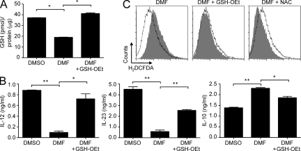
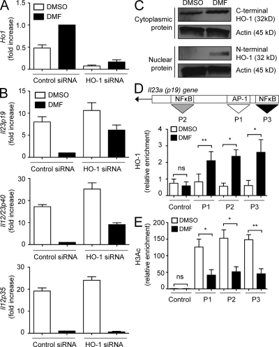
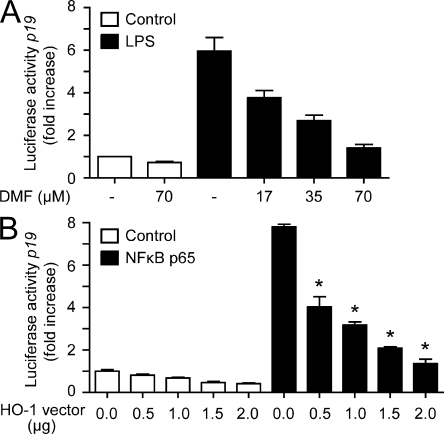
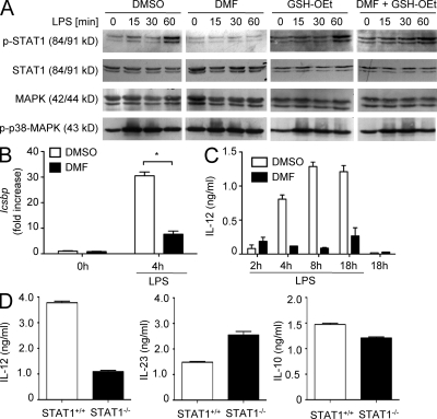
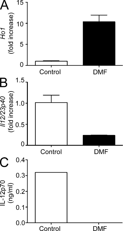
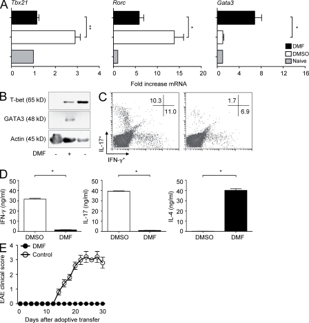
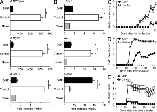
Fumarates improve multiple sclerosis (MS) and psoriasis, two diseases in which both IL-12 and IL-23 promote pathogenic T helper (Th) cell differentiation. However, both diseases show opposing responses to most established therapies. First, we show in humans that fumarate treatment induces IL-4-producing Th2 cells in vivo and generates type II dendritic cells (DCs) that produce IL-10 instead of IL-12 and IL-23. In mice, fumarates also generate type II DCs that induce IL-4-producing Th2 cells in vitro and in vivo and protect mice from experimental autoimmune encephalomyelitis. Type II DCs result from fumarate-induced glutathione (GSH) depletion, followed by increased hemoxygenase-1 (HO-1) expression and impaired STAT1 phosphorylation. Induced HO-1 is cleaved, whereupon the N-terminal fragment of HO-1 translocates into the nucleus and interacts with AP-1 and NF-κB sites of the IL-23p19 promoter. This interaction prevents IL-23p19 transcription without affecting IL-12p35, whereas STAT1 inactivation prevents IL-12p35 transcription without affecting IL-23p19. As a consequence, GSH depletion by small molecules such as fumarates induces type II DCs in mice and in humans that ameliorate inflammatory autoimmune diseases. This therapeutic approach improves Th1- and Th17-mediated autoimmune diseases such as psoriasis and MS by interfering with IL-12 and IL-23 production.


Figures

Figure 1.
Figure 1.
DMF therapy induces Th2 responses in human CD4+ T cells in vivo and human type II DCs. (A) CD4+ T were isolated from patients with psoriasis treated with either DMF or placebo at the indicated time of therapy and treated with PMA and ionomycin. IL-2, IL-4, and IFN-γ were assessed by intracellular flow cytometry. Each data point represents an individual patient sample (*, P < 0.05; **, P < 0.01). Horizontal bars represent the mean. (B) Quantification of intracellular GSH content of DCs treated with DMSO, 70 µM DMF, or 70 µM DMF and 1 mM GSH-OEt. Results are pooled data from four human donors (mean ± SEM; **, P < 0.01). (C–E) Human DCs were incubated with DMSO, 70 µM DMF, or DMF + 1 mM GSH-OEt and then treated with LPS for 18 h. IL-12, IL-23, or IL-10 production was determined by ELISA. Data were pooled from three donors (mean ± SEM; *, P < 0.05; **, P < 0.01).
Figure 2.
Figure 2.
DMF induces mouse type II macrophages and type II DCs. (A) BMDCs were incubated with DMSO, 70 µM DMF, or DMF + 1 mM GSH-OEt, and GSH content was determined by a colorimetric assay (mean ± SEM; *, P < 0.001). (B) DCs were treated with DMSO, 70 µM DMF, or DMF + 1 mM GSH-OEt and then stimulated with LPS for 18 h. Culture supernatants were harvested, and the indicated cytokines were determined by ELISA (mean ± SEM; *, P < 0.01; **, P < 0.001). (C) DCs were incubated with DMSO, 70 µM DMF, DMF + 1 mM GSH-OEt, or DMF and 1 mM NAC for 2–4 h, and intracellular ROS levels were assessed by staining with 2’,7’dichlorofluorescein. Intracellular ROS, gray; DMSO-treated controls, open. One representative experiment of three is shown.
Figure 3.
Figure 3.
DMF-induced HO-1 selectively prevents IL-23 induction. (A) DCs were treated with DMSO or 70 µM DMF, and HO-1 mRNA expression was determined by quantitative RT-PCR. HO-1 data were normalized to β-actin, and HO-1 level in control siRNA–transfected DMF–treated DCs was set as 1.0. The results are representative of three independent experiments. Error bars represent SEM. (B) HO-1 was knocked down, and levels of IL-12/IL-23p40, IL-23p19, or IL-12p35 mRNA were determined by RT-PCR. Data (mean ± SEM) were normalized to β-actin, and message levels in control siRNA–transfected DMF-treated DCs were set as 1.0. (C) DCs were treated as in A and lysed, and nuclear or cytoplasmic cell extracts were analyzed by Western blotting using antibodies directed against C- or N-terminal HO-1 protein. (D and E) DCs treated as in A were activated with LPS, cross-linked, and immunoprecipitated with anti–HO-1 (D) or anti-H3Ac (E). Bound DNA was amplified by quantitative PCR for primer sites P1 (AP-1; position 412–422 bp), P2 (c-Rel; position 560–584 bp), and P3 (RelA/c-Rel; position 394–406 bp). Data were pooled from four separate experiments and represent mean ± SEM (*, P < 0.05; **, P < 0.01; ns, not significant).
Figure 4.
Figure 4.
Interaction of DMF-induced HO-1 with the NF-κB p65-binding site directly inhibits IL-23p19 promoter activity. (A) RAW246.7 cells were transfected with a reporter construct containing the IL-23p19 promoter and treated with the indicated doses of DMF. Luciferase activity was measured after stimulation with 100 ng/ml LPS for 6 h. Data from one representative of two independent experiments are shown. Error bars represent SEM. (B) NIH 3T3 cells were transfected with a reporter construct containing the IL-23p19 promoter (1 µg/well) alone or together with the expression vector for p65 (1 µg/well), as well as the HO-1 or the empty control vector at the indicated concentration. Pooled data from two separate experiments with duplicates are shown (mean ± SEM; *, P < 0.001 relative to the empty vector control). Reporter gene data were normalized to the activity of cotransfected β-galactosidase.
Figure 5.
Figure 5.
DMF treatment selectively impairs IL-12 induction through inhibition of the p-STAT1–ICSBP–IL-12 signaling pathway. (A) DCs were treated with DMSO, 70 µM DMF, 1 mM GSH-OEt, or DMF + GSH-OEt and stimulated for the indicated time with LPS. STAT1, p-STAT1, MAPK, and p-p38 MAPK content of cell lysates was analyzed by Western blot. (B) DCs were incubated with DMSO or DMF and activated with LPS for 4 h. ICSBP expression was assessed by RT-PCR, and data were normalized to β-actin and expressed as fold increase (mean ± SEM; *, P < 0.01). ICSBP level in unstimulated cells (0 h) was set as 1.0. (C) DCs were incubated with DMSO or 70 µM DMF and stimulated with LPS for the indicated times. IL-12 levels in the culture supernatants were analyzed by ELISA. Error bars represent SEM. (D) DCs from STAT1+/+ or STAT1−/− mice were stimulated for 18 h with LPS. The indicated cytokines in culture supernatants were analyzed by ELISA. Data from one out of four experiments with similar results are shown (mean ± SEM).
Figure 6.
Figure 6.
Fumarate treatment induces HO-1 in DCs and inhibits IL-12/IL-23p40 production by DCs in vivo. (A–C) Mice were treated for 5 d with DMF in water or received DMF-free water (control). DCs were isolated, positively sorted, and stimulated with LPS for 18 h before analyzing HO-1 mRNA (A) or IL-12/IL-23p40 mRNA (B) expression by quantitative RT-PCR. RT-PCR data were normalized to β-actin levels, and expression in control mice was set as 1.0 (open bars). (C) DC isolated from DMF-treated or control mice were stimulated with LPS for 18 h, and IL-12p70 production was determined by ELISA. All data are shown as mean ± SEM and are representative for four independent experiments.
Figure 7.
Figure 7.
Type II DCs resulting from fumarate treatment selectively induce Th2 cells in vitro. (A) Naive OVA-specific CD4+ T cells were left unstimulated (gray bars) or were primed in vitro with OVA peptide and LPS-activated, DMSO-treated (open bars), or DMF-treated (black bars) DCs. Expression of the indicated transcription factors was assessed by RT-PCR, and the data were normalized to β-actin. Expression levels in unstimulated T cells were set at 1.0 (gray bars). Data are representative of three independent experiments (mean ± SEM; *, P < 0.05; **, P < 0.01). (B) DCs were treated and activated as in A and incubated alone (left lane) or with OVA-specific CD4+ T cells (middle and right lanes). T-bet or GATA3 protein expression in cell extracts was analyzed by Western blotting. (C and D) Naive OVA-specific CD4+ T cells were primed in vitro with OVA peptide and LPS-activated APCs in the presence or absence of DMF. Cells were expanded for 1 wk and then restimulated with OVA peptide and fresh APCs. Cytokines were determined by intracellular cytokine staining and flow cytometry (C) or by ELISA (D). Data from one representative experiment of three are shown (mean ± SEM; *, P < 0.001). (E) CD4+ T cells from immunized SJL mice were primed in vitro with PLP139-151 and APCs in the presence of DMF or DMSO (control) for 1 wk, restimulated, and expanded. 107 T cells were transferred into syngeneic WT mice (n = 5 per group), and EAE scores were determined. Data from one representative experiment of three are shown. Error bars represent SEM.
Figure 8.
Figure 8.
Fumarates induce type II APCs and Th2 cells in vivo and abolish the capacity of autoreactive CD4+ T cells to induce EAE. (A and B) SJL mice received DMF or DMF-free (control) water and were immunized with PLP139-151 peptide in CFA. On days 1–3, draining lymph nodes were isolated, RNA was extracted, and IL-12/IL-23p40, IL-12p35, or IL-23p19 expression (A) or Tbx21, Rorc, or Gata3 expression (B) was analyzed by RT-PCR (mean ± SEM; **, P < 0.001). Data were normalized to β-actin levels, and expression in naive mice was set as 1.0 (gray bars). Data from one representative experiment of three are shown (*, P < 0.01). (C) SJL mice were fed with DMF-containing or -free (control) water and immunized with PLP139-151 peptide in CFA and pertussis toxin (n = 5 per group). Clinical EAE scores were determined at the indicated times after immunization. Data are from one representative experiment of four. (D) TCR Vβ8.2 transgenic B10.PL mice were fed with 5 mg MMF or MMF-free (control) water (n = 8 per group). Mice were then immunized with MBP Ac1-11 peptide in CFA, and clinical scores were assessed at the indicated times after immunization. EAE incidence was 8/8 for control mice and 6/8 in the MMF group. (E) CD4+ T cells were isolated from spleens of MMF-treated or control donors from D on day 42, and equal numbers of cells (107) were adoptively transferred into naive mice. EAE scores were assessed at the indicated times after adoptive transfer (MMF group, n = 4; control group, n = 6). Experiments with MMF were performed four times in B10.PL or SJL mice, and data shown are from one representative experiment. (C and E) Error bars represent SEM.

References

    1. Altmeyer P.J., Matthes U., Pawlak F., Hoffmann K., Frosch P.J., Ruppert P., Wassilew S.W., Horn T., Kreysel H.W., Lutz G., et al. 1994. Antipsoriatic effect of fumaric acid derivatives. Results of a multicenter double-blind study in 100 patients. J. Am. Acad. Dermatol. 30:977–981 10.1016/S0190-9622(94)70121-0 - DOI - PubMed
    1. Arbiser J.L. 2011. Fumarate esters as angiogenesis inhibitors: key to action in psoriasis? J. Invest. Dermatol. 131:1189–1191 10.1038/jid.2011.45 - DOI - PubMed
    1. Axtell R.C., de Jong B.A., Boniface K., van der Voort L.F., Bhat R., De Sarno P., Naves R., Han M., Zhong F., Castellanos J.G., et al. 2010. T helper type 1 and 17 cells determine efficacy of interferon-beta in multiple sclerosis and experimental encephalomyelitis. Nat. Med. 16:406–412 10.1038/nm.2110 - DOI - PMC - PubMed
    1. Becher B., Durell B.G., Noelle R.J. 2002. Experimental autoimmune encephalitis and inflammation in the absence of interleukin-12. J. Clin. Invest. 110:493–497 - PMC - PubMed
    1. Bettelli E., Sullivan B., Szabo S.J., Sobel R.A., Glimcher L.H., Kuchroo V.K. 2004. Loss of T-bet, but not STAT1, prevents the development of experimental autoimmune encephalomyelitis. J. Exp. Med. 200:79–87 10.1084/jem.20031819 - DOI - PMC - PubMed

Publication types

MeSH terms