. 2011 Dec 1;71(23):7198-206.
doi: 10.1158/0008-5472.CAN-11-1514. Epub 2011 Oct 10.

Chronic activation of wild-type epidermal growth factor receptor and loss of Cdkn2a cause mouse glioblastoma formation

Affiliations

Chronic activation of wild-type epidermal growth factor receptor and loss of Cdkn2a cause mouse glioblastoma formation

Jaime Acquaviva et al. Cancer Res. .

Abstract

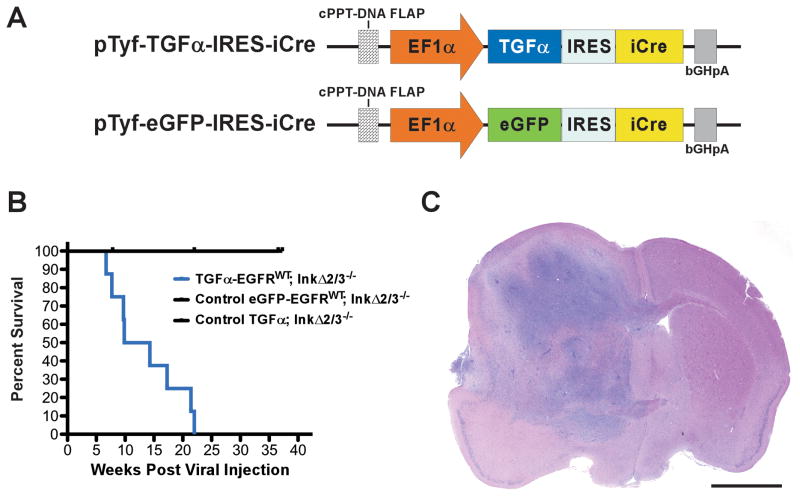
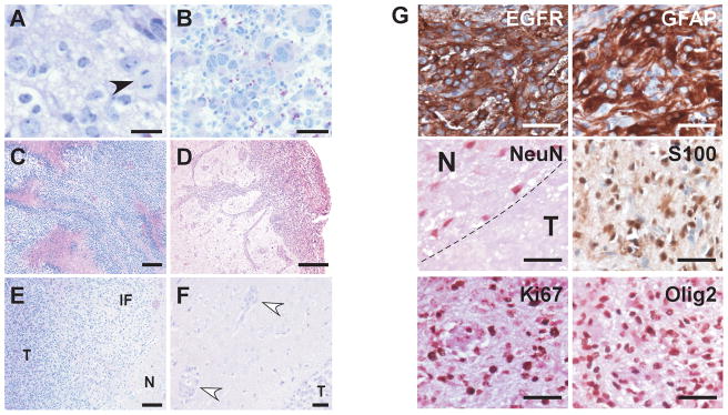
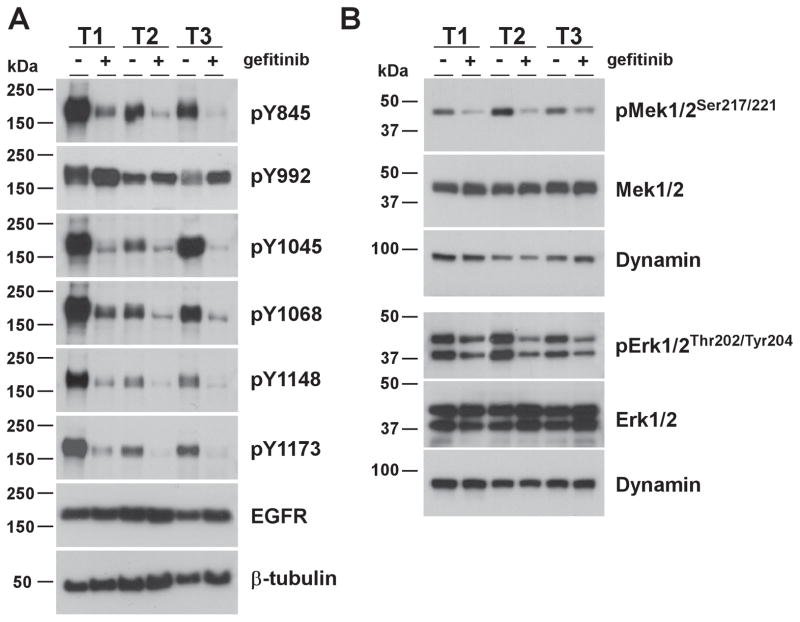
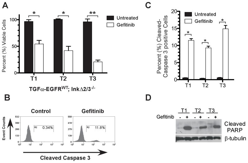
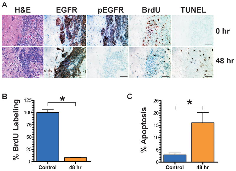
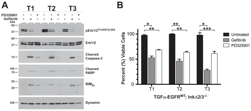
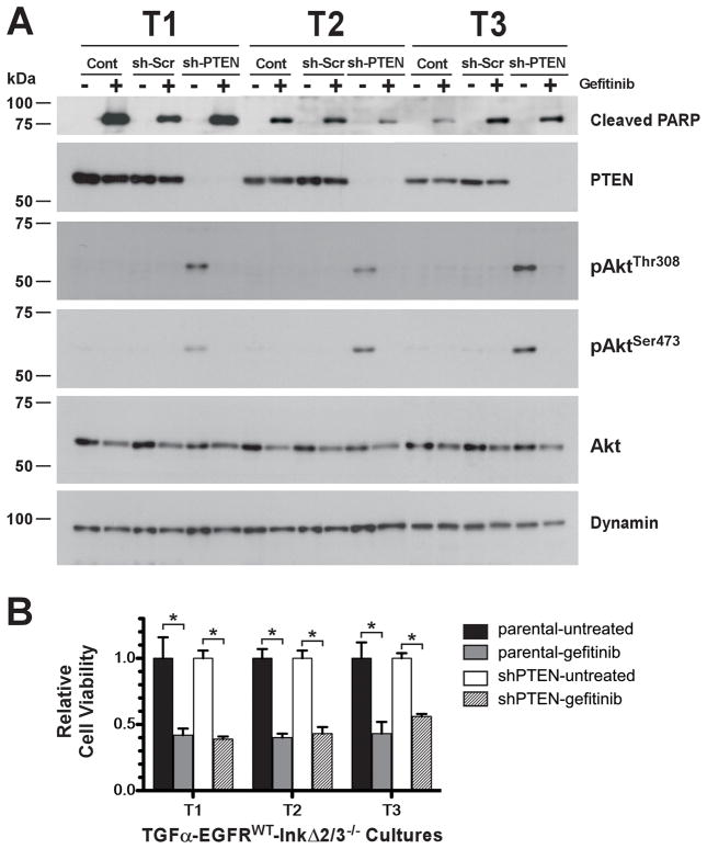
Glioblastoma multiforme (GBM) is characterized by overexpression of epidermal growth factor receptor (EGFR) and loss of the tumor suppressors Ink4a/Arf. Efforts at modeling GBM using wild-type EGFR in mice have proven unsuccessful. Here, we present a unique mouse model of wild-type EGFR-driven gliomagenesis. We used a combination of somatic conditional overexpression and ligand-mediated chronic activation of EGFR in cooperation with Ink4a/Arf loss in the central nervous system of adult mice to generate tumors with the histopathologic and molecular characteristics of human GBMs. Sustained, ligand-mediated activation of EGFR was necessary for gliomagenesis, functionally substantiating the clinical observation that EGFR-positive GBMs from patients express EGFR ligands. To gain a better understanding of the clinically disappointing EGFR-targeted therapies for GBM, we investigated the molecular responses to EGFR tyrosine kinase inhibitor (TKI) treatment in this model. Gefitinib treatment of primary GBM cells resulted in a robust apoptotic response, partially conveyed by mitogen-activated protein kinase (MAPK) signaling attenuation and accompanied by BIM(EL) expression. In human GBMs, loss-of-function mutations in the tumor suppressor PTEN are a common occurrence. Elimination of PTEN expression in GBM cells posttumor formation did not confer resistance to TKI treatment, showing that PTEN status in our model is not predictive. Together, these findings offer important mechanistic insights into the genetic determinants of EGFR gliomagenesis and sensitivity to TKIs and provide a robust discovery platform to better understand the molecular events that are associated with predictive markers of TKI therapy.


Conflict of interest statement

No potential conflicts of interest were disclosed.

Figures

Figure 1
Figure 1
Activated EGFRWT cooperates with loss of tumor suppressor genes to form brain tumors that have characteristics of GBM. A, Schematic representation of the pTyf lentiviral transducing vectors. This bicistronic vector is derived from a previously described self-inactivating virus (46) modified to contain the human TGFα cDNA followed by a poliovirus1 IRES and improved Cre (iCre) cDNA (50) driven by the human elongation factor-1α (EF1α) promoter. For control experiments, TGFα is replaced with the eGFP gene. The presence of a central polypurine tract (cPPT)-DNA FLAP element upstream of the multiple cloning site significantly improves the transduction efficiency in CNS tissues (48, 49). B, Survival (Kaplan-Meier) analysis of conditional EGFR mice. Cohorts of mice of the indicated genotypes were stereotactically injected in the striatum with titer-matched pTyf-TGFα-IRES-iCre or pTyf-eGFP-IRES-iCre and monitored for survival over time. C, Photomicrograph of an H&E-stained coronal section of a TGFα-EGFRWT;InkΔ2/3−/− brain tumor. Scale bar; 2.0 mm.
Figure 2
Figure 2
Representative histological photomicrographs of TGFα-EGFRWT;InkΔ2/3−/− tumors. H&E-stained paraffin-embedded tumor sections; A, Tumors are set on a fibrillary background and contain densely packed cells featuring pleimorphic nuclei with prominent nucleoli and mitoses (black arrowhead). B, Giant multinucleated cells are present within tumors. C, Tumors exhibit marked pseudopallisading necrosis. D–F, the highly infiltrative nature of TGFα-EGFRWT tumor cells is depicted, (D) tumor cells migrate within meninges in the subarachnoid space and invade the Virchow-Robin space and (E) are infiltrating normal parenchyma (N) by forming a loose infiltrating front (IF) away from the bulk tumor (T) and (F) tumor cells migrate along blood vessels and invade the perivascular space (white arrow head) distant from the bulk tumor (T). G, EGFRWT GBM tumors express markers of astrocytic differentiation. Representative photomicrographs of tumors stained with cell lineage markers using IHC. Tumors stain positive for markers of astrocytic lineage (glial fibrillary acidic protein (GFAP) and S100) and negative for markers of neuronal (NeuN) lineage. GBM tumors also stain positive for human EGFR, the proliferation marker Ki67, and for Olig2. EGFR, GFAP and S100 sections were counterstained with hematoxylin and sections for the nuclear NeuN, Olig2 and Ki67 markers were counterstained with eosin. N, normal brain; T, tumor. Scale bars; 25 μm (A), 50 μm (B,F,G), 62.5 μm (D), 125 μm (C, E).
Figure 3
Figure 3
EGFRWT kinase inhibition attenuates signaling pathways. A–B, Immunoblot of total cell extracts from vehicle- and gefitinib-treated (10 μM) TGFα-EGFRWT;InkΔ2/3−/−tumor cultures analyzed for the presence of (A) the indicated phosphotyrosine residues (B) the activation status of the canonical MAPK members Mek1/2 and Erk1/2.β-tubulin and dynamin are used as internal loading controls.
Figure 4
Figure 4
EGFR kinase inhibition in TGFα-EGFRWT;InkΔ2/3−/− GBM tumor cells is cytotoxic. A, Tumor cells are sensitive to gefitinib treatment. Viability assay of three independent tumor cell cultures (T1-3) after vehicle or gefitinib treatment (10 μM) for 24 hours. Data is plotted as percentage of viable cells of treated over mock treatment (mean ± SD; n=3 in each group, *p<0.005, **p<0.0005, two-tailed t-test). B, Representative flow-cytometric analysis and C, graphical representation of TGFα-EGFRWT;InkΔ2/3−/− GBM primary cell cultures mock- and gefitinib-treated indicating an increase in cleaved caspase-3 positive cells upon EGFR kinase inhibitor treatment (mean ± SD; n=3 in each group, *p<0.0001, two-tailed t-test). D, immunoblot of total cell extracts from vehicle- and gefitinib-treated (10 μM) cultures of the TGFα-EGFRWT;InkΔ2/3−/− GBM tumor cells analyzed for presence of the apoptotic marker cleaved PARP. β-tubulin is used as an internal loading control.
Figure 5
Figure 5
Orthotopic allograft tumors of TGFα-EGFRWT;InkΔ2/3−/− GBM cells are sensitive to EGFR inhibition. A, representative photomicrographs of paraffin-embedded tumor tissue sections stained for the indicated markers from control (0 hr) and treated tumor-bearing animals 48 hr post treatment. B, graphical representation of the quantification of proliferation assayed by BrdU incorporation. The BrdU staining data is presented as the percentage of BrdU positive cells in treated tumors over control tumors. C, graphical representation of the quantification of the percentage of apoptotic cells as measured by the number of TUNEL positive cells. Quantification of apoptosis is presented as percentage of TUNEL positive cells per field of view. (mean ± S.D., n=6 in each group *p=0.0001, two-tailed t-test). Scale bar: 250 μm.
Figure 6
Figure 6
EGFRWT inhibition-induced apoptosis is partly mediated by MAPK signaling attenuation. TGFα-EGFRWT;InkΔ2/3−/− GBM tumor cell cultures (T1-T3) were treated with gefitinib (10 μM) or Mek1/2 inhibitor PD325901 (100 nM) for 24 hr and A, analyzed by immunoblot analysis for the apoptotic markers cleaved caspase-3 and cleaved PARP and for the pro-apoptotic protein BIMEL and B, analyzed for viability in a growth assay. Data is plotted as percentage of viable cells of treated over mock treatment (mean ± SD; n=3 in each group, *p=0.0002, **p<0.0001, ***p<0.005, two-tailed t-test)..
Figure 7
Figure 7
PTEN loss does not confer resistance to EGFR kinase inhibition. A, cells expressing a scrambled control sh-RNA (sh-Scr) or a PTEN sh-RNA (sh-PTEN) were analyzed by immunoblot for cleaved PARP, phospho AktThr308, AktSer473 and PTEN expression. Total Akt and β-tubulin are used as an internal loading control. B, parental TGFα-EGFRWT;InkΔ2/3−/− cultured tumor cells (T1-T3) and their PTEN knockdown counterpart were treated with gefitinib (10 μM) and assayed for cell viability. The results are presented as values relative to untreated conditions (mean ± SD; n=3 in each group, *p<0.005, two-tailed t-test).

References

    1. Brennan C. Genomic profiles of glioma. Curr Neurol Neurosci Rep. 2011;11:291–7. - PubMed
    1. Furnari FB, Fenton T, Bachoo RM, Mukasa A, Stommel JM, Stegh A, et al. Malignant astrocytic glioma: genetics, biology, and paths to treatment. Genes Dev. 2007;21:2683–710. - PubMed
    1. Huang PH, Xu AM, White FM. Oncogenic EGFR signaling networks in glioma. Sci Signal. 2009;2:re6. - PubMed
    1. http://cancergenome.nih.gov.

    1. McLendon R, Friedman A, Bigner D, Van Meir EG, Brat DJ, Mastrogianakis M, et al. Comprehensive genomic characterization defines human glioblastoma genes and core pathways. Nature. 2008;455:1061–8. - PMC - PubMed

Publication types

MeSH terms