. 2011 Nov;204 Suppl 3(Suppl 3):S957-67.
doi: 10.1093/infdis/jir326.

Ebola virus enters host cells by macropinocytosis and clathrin-mediated endocytosis

Affiliations

Ebola virus enters host cells by macropinocytosis and clathrin-mediated endocytosis

Paulina Aleksandrowicz et al. J Infect Dis. 2011 Nov.

Abstract

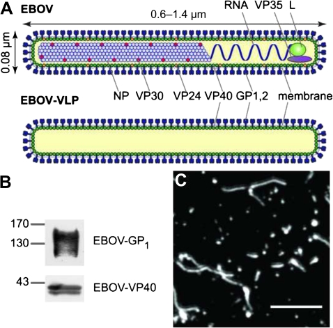
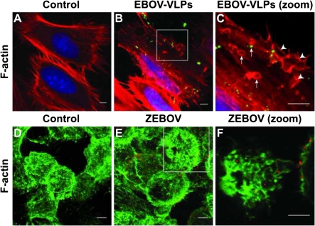
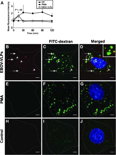
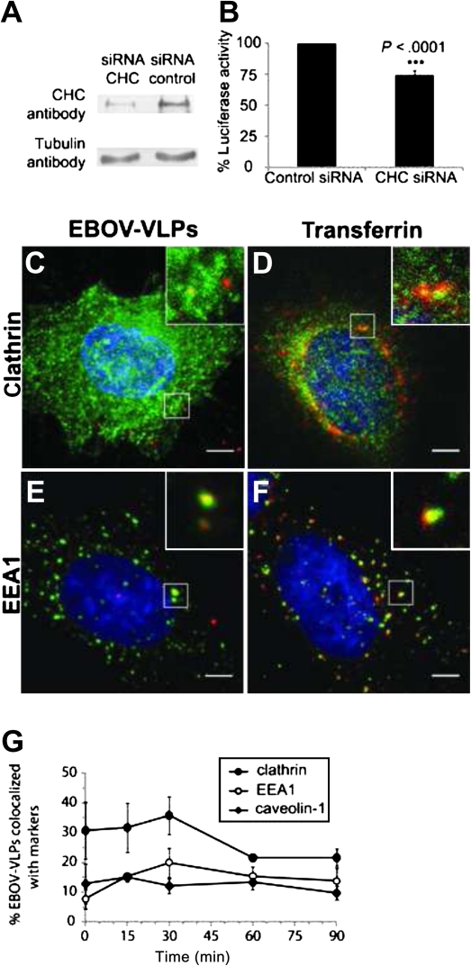
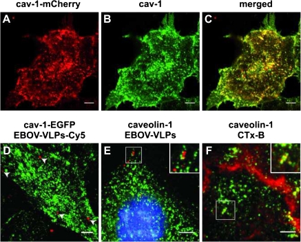
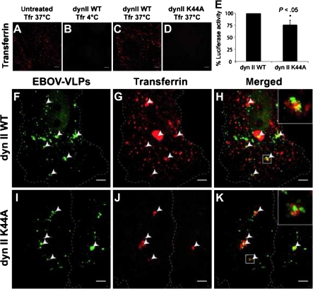
Virus entry into host cells is the first step of infection and a crucial determinant of pathogenicity. Here we show that Ebola virus-like particles (EBOV-VLPs) composed of the glycoprotein GP(1,2) and the matrix protein VP40 use macropinocytosis and clathrin-mediated endocytosis to enter cells. EBOV-VLPs applied to host cells induced actin-driven ruffling and enhanced FITC-dextran uptake, which indicated macropinocytosis as the main entry mechanism. This was further supported by inhibition of entry through inhibitors of actin polymerization (latrunculin A), Na(+)/H(+)-exchanger (EIPA), and PI3-kinase (wortmannin). A fraction of EBOV-VLPs, however, colocalized with clathrin heavy chain (CHC), and VLP uptake was reduced by CHC small interfering RNA transfection and expression of the dominant negative dynamin II-K44A mutant. In contrast, we found no evidence that EBOV-VLPs enter cells via caveolae. This work identifies macropinocytosis as the major, and clathrin-dependent endocytosis as an alternative, entry route for EBOV particles. Therefore, EBOV seems to utilize different entry pathways depending on both cell type and virus particle size.


Figures

Figure 1.
Figure 1.
Ebola viruses (EBOVs) and Ebola virus-like particles (EBOV-VLPs). A, A scheme of EBOV and EBOV-VLPs illustrating the overall organization. The minimal components of EBOV-VLPs are the glycoprotein GP1,2 on the surface and the matrix protein VP40, which is needed for particle formation. B, Western blot analysis of EBOV-VLPs showing the presence of GP1, the larger cleavage fragment, and VP40. C, EBOV-VLPs stained for EBOV-GP1,2. EBOV-VLPs are similar in morphology to infectious EBOV. Scale bar: 2 μm.
Figure 2.
Figure 2.
Ebola virus-like particles (EBOV-VLPs) and Zaire ebolavirus (ZEBOV) induce formation of actin ruffles and macropinosomes. A–C, HeLa cells were exposed to EBOV-VLPs or phosphate-buffered saline (control) for 30 minutes, and fixed with formaldehyde. Actin filaments were labeled with phalloidin-TRITC (red) and EBOV-VLPs with a GP1,2-specific antibody (green). Higher magnification of the boxed area reveals actin ruffles (arrowheads) and macropinosomes (arrows) with and without EBOV-VLPs. D–F, Formaldehyde-fixed HeLa cells, 15 minutes after exposure to ZEBOV (multiplicity of infection = 1), were stained for filamentous actin with phalloidin–fluorescein isothiocyanate (FITC) (green) and for EBOV with antibody against EBOV-VP40 (magenta). Higher magnification of the boxed area reveals virus particles engulfed by actin ruffles and inside macropinosomes. Experiments were repeated 3 times with similar results.
Figure 3.
Figure 3.
Ebola virus-like particles (EBOV-VLPs) stimulate fluid-phase macropinocytosis in Vero and HeLa cells. A, EBOV-VLPs were bound to Vero E6 cells on ice, shifted to 37°C for the indicated time periods, exposed for fluorescein isothiocyanate (FITC)–dextran during the last 10 minutes, and analyzed by fluorescence-activated cell sorting. Phorbol 12-myristate 13-acetate (PMA) (200 nM) served as a positive control. EBOV-VLPs increased the FITC-dextran uptake transiently. B–D, Addition of EBOV-VLPs-Cy5 (red) and FITC-dextran (green); or E–G, 200 nM PMA and FITC-dextran (green); or H–J, only FITC-dextran (green) to HeLa cells 30 minutes after temperature shift to 37°C. Both EBOV-VLPs-Cy5 and PMA (used as positive control) stimulate uptake of fluid phase as indicated by FITC-dextran-positive macropinosomes. Dotted lines indicate cell borders. FITC-dextran (green); EBOV-VLP-Cy5 (red); nuclei labeled with DAPI (blue). Experiment was repeated 4 times. Bars in A represent standard errors. Scale bar: 5 μm.
Figure 4.
Figure 4.
Inhibitors of macropinocytosis block Ebola infectious virus-like particle (EBOV-iVLP) infection and uptake of recombinant Ebola virus (EBOV). HeLa cells and Vero E6 cells were treated with 5-(N-Ethyl-N-isopropyl)-amiloride (EIPA) (up to 200 μM), latrunculin A (0.5 μM), or 100 nM wortmannin prior to infection. A, HeLa cells exposed to EIPA were infected with EBOV-iVLPs leading to the expression of Renilla luciferase, which was dose-dependently inhibited by EIPA. B and C, EIPA, latrunculin A, and wortmannin decreased EBOV-iVLP infection in both cell types. D and E, Vero E6 cells were treated with either EIPA or latrunculin as indicated and subsequently infected with infectious Zaire ebolavirus–green fluorescent protein (ZEBOV-GFP). Inhibition of enhanced green fluorescent protein (EGFP) expression was seen for EIPA and latrunculin. Each experiment with EBOV-iVLPs was repeated 4–7 times. Experiments with infectious EBOV-EGFP were repeated 3 times. Error bars represent standard errors. P values indicate significance. DMSO, dimethyl sulfoxide.
Figure 5.
Figure 5.
Clathrin modulates Ebola infectious virus-like particle (EBOV-iVLP) entry into HeLa cells. A, Downregulation of clathrin heavy chain (CHC) in HeLa cells by small interfering RNA (siRNA), as shown by Western blotting. B, Clathrin-depleted HeLa cells and control cells were infected with EBOV–iVLPs that mediate expression of Renilla luciferase, which was used as readout for infectivity. Despite a 75% downregulated CHC, the reduction of EBOV-iVLP infection was only 25%. C and D, Virus-like particle (VLP) adsorption was performed for 1 hour at 4°C followed by a 15-minute temperature shift to 37°C. Colocalization of VLPs (stained with anti-EBOV-VP40 antibody) with either clathrin or early endosome antigen 1 (EEA1), and transferrin with clathrin or EEA1 was determined by immunofluorescence analysis. G, Quantification of VLPs colocalized with clathrin, EEA1, or caveolin-1 at indicated time points. Data are based on 3 independent experiments. Error bars represent standard errors. Scale bar: 5 μm.
Figure 6.
Figure 6.
Ebola virus-like particles (EBOV-VLPs) do not colocalize with caveolin-1 in HeLa cells. A, Fluorescent caveolin-1-mCherry (red) was expressed in HeLa cells by lentiviral gene transfer. B, Caveolin-1 was labeled using an anti-caveolin-1 antibody and Alexa Fluor 488–conjugated secondary antibody (green). C, The merged image shows colocalization of caveolin-1 and the additionally expressed caveolin-1-mCherry. D, Caveolin-1–enhanced green fluorescent protein (EGFP)-expressing HeLa cells were exposed to EBOV-VLPs-Cy5 at 4°C followed by a temperature shift to 37°C. Live cell imaging was performed, and time-dependent 3-dimensional stacks were acquired followed by 3-dimensional reconstruction. No colocalization of EBOV-VLPs (red) or caveolin-1-EGFP (green) was observed. E, HeLa cells were exposed to EBOV-VLPs for 1 hour at 4°C followed by a 15-minute temperature shift to 37°C, fixed, and stained for caveolin-1 and EBOV-VLPs using an antibody directed against GP1,2. Most of the VLPs were not colocalized (insert, red), but a very few VLPs overlapped with cav-1 staining (insert, yellow) F, Cholera toxin B (CTx-B) served as a positive control and colocalized with caveolin-1. Scale bar: 5 μm.
Figure 7.
Figure 7.
Dynamin II modulates Ebola virus-like particle (EBOV-VLP) uptake in HeLa cells. Dynamin II-WT (dynII WT) and a dynamin II-K44A mutant (dynII K44A) were expressed in HeLa cells to block both caveolin-1 and clathrin-mediated endocytosis, and subsequently tested for the ability to take up EBOV-VLPs, or transferrin (positive control). A–D, HeLa cells expressing either dynamin II-WT or dynamin II-K44A mutant were exposed to transferrin–Alexa Fluor 546 at temperatures indicated. There was no transferrin uptake in cells expressing the dynamin II K44A mutant. E, HeLa cells expressing either dynamin II or dynamin II-K44A were infected with EBOV-iVLPs and analyzed for Renilla luciferase activity after 70 hours. There was 25% downregulation of luciferase activity in dynamin II-K44A–expressing cells. F–K, HeLa cells expressing dynamin II-WT (F–H) or dynamin II K44A mutant (I–K) were exposed to EBOV-VLPs for 60 minutes and to transferrin–Alexa Fluor 546 for 15 minutes and fixed, and EBOV-VLPs (green) were stained for GP1,2. F–H, Some EBOV-VLPs were found in exceptionally large transferrin-positive vesicles, while the bulk of EBOV-VLPs did not colocalize with transferrin. I–K, In contrast, HeLa cells expressing dynamin II-K44A were negative for the characteristic small transferrin-positive vesicles, but still showed the exceptionally large EBOV-VLP–containing macropinosomes. Arrowheads indicate colocalized structures as indicated. Experiment was repeated 3 times. Error bars represent standard errors. Scale bars: 5 μm.

References

    1. Sanchez A, Geisbert TW, Feldmann H. Marburg and Ebola viruses. In: Fields BN, Knipe DM, Howley PM, editors. Fields virology. 5th ed. Philadelphia: Wolters Kluwer Health/Lippincott Williams & Wilkins; 2007.
    1. Feldmann H, Jones S, Klenk HD, Schnittler HJ. Ebola virus: from discovery to vaccine. Nat Rev Immunol. 2003;3:677–85. - PubMed
    1. Geisbert TW, Jahrling PB. Exotic emerging viral diseases: progress and challenges. Nat Med. 2004;10:S110–21. - PubMed
    1. Schnittler HJ, Feldmann H. Marburg and Ebola hemorrhagic fevers: does the primary course of infection depend on the accessibility of organ-specific macrophages? Clin Infect Dis. 1998;27:404–6. - PubMed
    1. Feldmann H, Volchkov VE, Volchkova VA, Stroher U, Klenk HD. Biosynthesis and role of filoviral glycoproteins. J Gen Virol. 2001;82:2839–48. - PubMed

Publication types

MeSH terms