. 2011 Oct 18;108(42):17372-7.
doi: 10.1073/pnas.1114465108. Epub 2011 Oct 10.

Autoantibodies recognizing carbamylated proteins are present in sera of patients with rheumatoid arthritis and predict joint damage

Affiliations

Autoantibodies recognizing carbamylated proteins are present in sera of patients with rheumatoid arthritis and predict joint damage

Jing Shi et al. Proc Natl Acad Sci U S A. .

Abstract

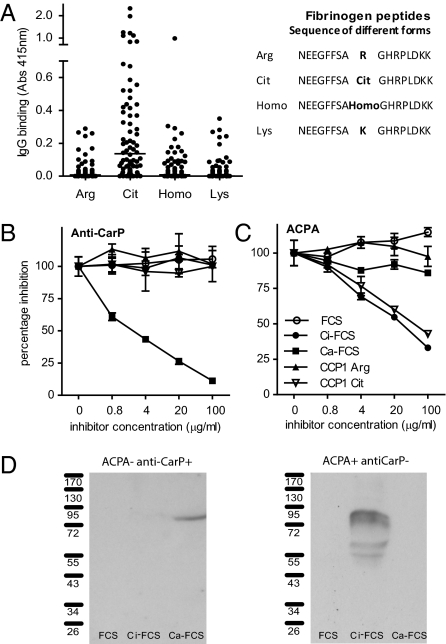
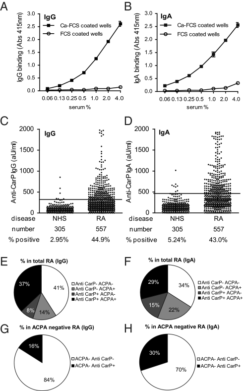
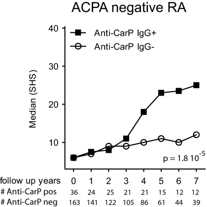
Autoimmune responses against posttranslationally modified antigens are a hallmark of several autoimmune diseases. For example, antibodies against citrullinated protein antigens (ACPA) have shown their relevance for the prognosis and diagnosis of rheumatoid arthritis (RA), and have been implicated in disease pathogenesis. It is conceivable that other autoantibody systems, recognizing other posttranslationally modified proteins, are also present in RA. Here, we describe the presence of an autoantibody system that discriminates between citrulline- and homocitrulline-containing antigens in the sera of RA-patients. IgG antibodies recognizing carbamylated (homocitrulline-containing) antigens were present in sera of over 45% of RA-patients. Likewise, anticarbamylated protein (anti-CarP) IgA antibodies were observed in 43% of RA-sera. ACPA and anti-CarP antibodies are distinct autoantibodies because, in selected double-positive patients, the anti-CarP antibody binding to carbamylated antigens could be inhibited by carbamylated antigens, but not by control or citrullinated antigens. Similarly, ACPA-binding to citrullinated antigens could only be inhibited by citrullinated antigens. In line with this observation, 16% of ACPA-negative RA-patients, as measured by a standard ACPA assay, harbored IgG anti-CarP antibodies, whereas 30% of these patients tested positive for IgA anti-CarP antibodies. The presence of anti-CarP antibodies was predictive for a more severe disease course in ACPA-negative patients as measured by radiological progression. Taken together, these data show the presence of a unique autoantibody system recognizing carbamylated, but not citrullinated, protein antigens. These antibodies are predictive for a more severe clinical course in ACPA-negative RA-patients, indicating that anti-CarP antibodies are a unique and relevant serological marker for ACPA-negative RA.


Conflict of interest statement

The authors declare no conflict of interest.

Figures

Fig. 1.
Fig. 1.
Illustration of citrullination and carbamylation. Citrullination (A) and carbamylation (B) occur on different amino acids via different mechanisms, but yield similar end-products.
Fig. 2.
Fig. 2.
Antibodies against carbamylated proteins are present in sera of RA patients. The reactivity of IgG (A and B) and IgA (D and E) from sera of healthy controls (NHS) or RA patients (RA) to wells coated with nonmodified FCS (FCS) or carbamylated FCS (Ca-FCS) is depicted. Data expressed as absorbance at 415 nm. (C and F) Absorbance units of FCS were subtracted from the absorbance units of Ca-FCS, representing the specific anticarbamylated protein response. ***P < 0.0001 for a t test comparing NHS and RA.
Fig. 3.
Fig. 3.
Anti-CarP antibodies and ACPA are two separate autoantibody systems. (A) IgG reactivity of 76 sera from RA patients, toward several forms of a Fib peptide is depicted. (B and C) Antibody binding to Ca-FCS or Ci-FCS was inhibited using preincubations with fluid-phase inhibitors. (D) FCS, Ca-FCS, and Ci-FCS were separated by SDS-PAGE gels and blotted. The presence of antibodies reactive to proteins on the blots was analyzed by incubating these blots with either anti-CarP–positive ACPA-negative and anti-CarP–negative ACPA-positive sera.
Fig. 4.
Fig. 4.
Anti-CarP antibodies bind to Ca-Fib via variable domains. (A) IgG reactivity against Fib, Ci-Fib, and Ca-Fib of 54 healthy controls and 214 RA patients was analyzed by ELISA. (B) Specificity of anti–Ca-Fib reactivity was confirmed using inhibition studies. One sample is shown, where data are expressed relative to inhibition with PBS. (C) The molecular nature of purified IgG and F(ab′)2 was confimed by Coomassie-stained SDS-PAGE gel. (D) F(ab′)2 fragments were generated from purified IgG of 2 anti-CarP–positive patients and two negative controls. Only F(ab′)2 from patients reacted with Ci-Fib and Ca-Fib. (E) Inhibition experiments confirm that also F(ab′)2 are not necessarily cross-reactive between Ci-Fib and Ca-Fib.
Fig. 5.
Fig. 5.
Anti-CarP IgG and IgA antibodies are present in RA sera. (A and B) Dose-response curves of the anti-CarP antibody-positive standard (IgG and IgA) on Ca-FCS and FCS in ELISA. (C and D) ELISA was performed for the detection of anti-CarP IgG and IgA in sera of healthy controls (NHS) and RA patients. A cut-off was established using the mean plus two times the SD of the healthy controls, as described in the Materials and Methods. Reactivity is depicted as arbitrary units per milliliter. The number of samples tested and the percentage of positivity is indicated below the graph. (E and F) Pie charts showing the percentage of RA patients positive and negative for anti-CCP2 and anti-CarP antibodies. (G and H) Pie charts showing the percentage of anti-CarP IgG- or IgA-positive patients negative for anti-CCP2.
Fig. 6.
Fig. 6.
Anti-CarP IgG antibodies are associated with a more severe radiological progression in ACPA-negative RA. The extent and rate of joint destruction were analyzed in all RA patients included, or analyzed separately, for ACPA-negative or ACPA-positive subgroups (Fig. S1). The severity of joint destruction is depicted as median Sharp–van der Heijde score (SHS) on the y axis and the follow-up years on the x axis. Below the x axis, the patient number is listed for each time point. Radiological progression for the anti-CCP2–negative RA patients is shown. The P value is derived from the analysis model, as described in Materials and Methods.

Similar articles

Cited by

References

    1. Klareskog L, Rönnelid J, Lundberg K, Padyukov L, Alfredsson L. Immunity to citrullinated proteins in rheumatoid arthritis. Annu Rev Immunol. 2008;26:651–675. - PubMed
    1. Kallberg H, et al. Epidemiological Investigation of Rheumatoid Arthritis study group Gene-gene and gene-environment interactions involving HLA-DRB1, PTPN22, and smoking in two subsets of rheumatoid arthritis. Am J Hum Genet. 2007;80:867–875. - PMC - PubMed
    1. van der Helm-van Mil AH, Verpoort KN, Breedveld FC, Toes RE, Huizinga TW. Antibodies to citrullinated proteins and differences in clinical progression of rheumatoid arthritis. Arthritis Res Ther. 2005;7:R949–R958. - PMC - PubMed
    1. van der Woude D, et al. Prevalence of and predictive factors for sustained disease-modifying antirheumatic drug-free remission in rheumatoid arthritis: Results from two large early arthritis cohorts. Arthritis Rheum. 2009;60:2262–2271. - PubMed
    1. Visser K, et al. Pretreatment serum levels of anti-cyclic citrullinated peptide antibodies are associated with the response to methotrexate in recent-onset arthritis. Ann Rheum Dis. 2008;67:1194–1195. - PubMed

Publication types