. 2012 Apr 10;21(6):846-58.
doi: 10.1089/scd.2011.0150. Epub 2011 Oct 11.

Conversion of human bone marrow-derived mesenchymal stem cells into tendon progenitor cells by ectopic expression of scleraxis

Affiliations

Conversion of human bone marrow-derived mesenchymal stem cells into tendon progenitor cells by ectopic expression of scleraxis

Paolo Alberton et al. Stem Cells Dev. .

Abstract

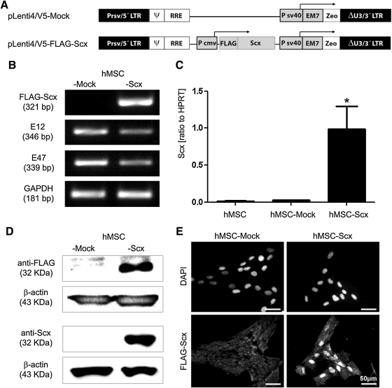
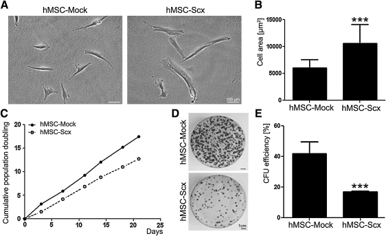
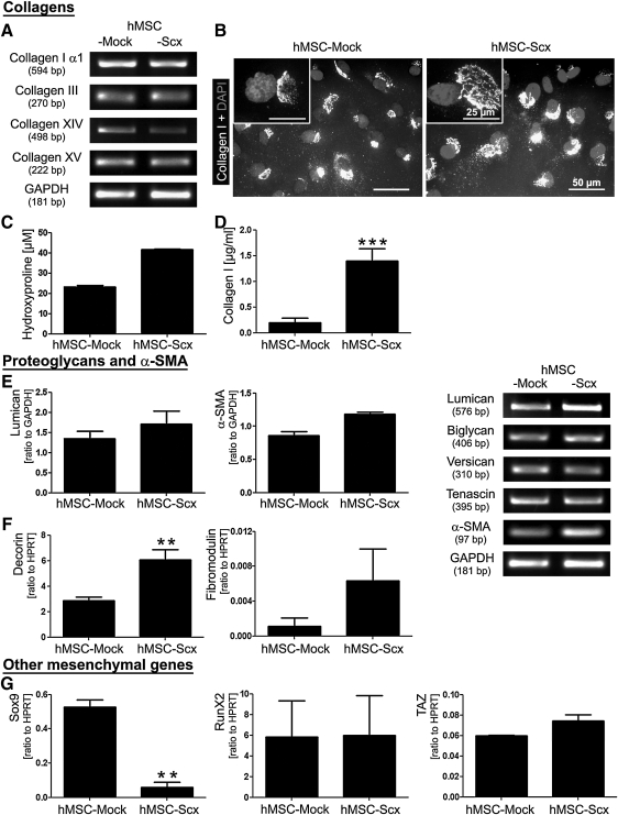
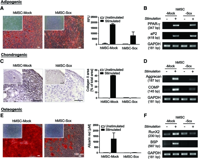
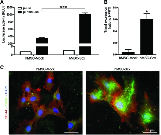
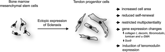
Tendons and ligaments (T/L) are dense connective tissues of mesodermal origin. During embryonic development, the tendon-specific cells descend from a sub-set of mesenchymal progenitors condensed in the syndetome, a dorsolateral domain of the sclerotome. These cells are defined by the expression of the transcription factor scleraxis (Scx), which regulates tendon formation and several other characteristic genes, such as collagen type I, decorin, fibromodulin, and tenomodulin (Tnmd). In contrast to other mesenchymal progenitors, the genealogy and biology of the tenogenic lineage is not yet fully understood due to the lack of simple and efficient protocols enabling generation of progenitors in vitro. Here, we investigated whether the expression of Scx can lead to the direct commitment of mesenchymal stem cells (MSCs) into tendon progenitors. First, MSC derived from human bone marrow (hMSC) were lentivirally transduced with FLAG-Scx cDNA to establish 2 clonal cell lines, hMSC-Scx and hMSC-Mock. Subsequent to Scx transduction, hMSC underwent cell morphology change and had significantly reduced proliferation and clonogenicity. Gene expression analysis demonstrated that collagen type I and several T/L-related proteoglycans were upregulated in hMSC-Scx cells. When stimulated toward 3 different mesenchymal lineages, hMSC-Scx cells failed to differentiate into chondrocytes and osteoblasts, whereas adipogenic differentiation still occurred. Lastly, we detected a remarkable upregulation of the T/L differentiation gene Tnmd in hMSC-Scx. From these results, we conclude that Scx delivery results in the direct programming of hMSC into tendon progenitors and that the newly generated hMSC-Scx cell line can be a powerful and useful tool in T/L research.


Figures

FIG. 1.
FIG. 1.
Lentiviral transduction of FLAG-Scx cDNA in hMSC and establishment of stable cell lines. (A) Plasmid charts of pLenti/V5-FLAG-Scx expression construct and control, Mock construct. (B) Semi-quantitative RT-PCR analysis for FLAG-Scx transgene (primer set annealing on the FLAG and Scx cDNA) and E12 and E47 Scx-dimerization partners. (C) Scx quantitative RT-PCR with a primer set annealing within Scx cDNA. GAPDH and HPRT were used as reference genes, and all RT-PCR results were reproduced at least twice independently. Bar charts present mean±standard deviation; *P<0.05. (D) Western blot analysis of hMSC-Mock and hMSC-Scx cells with anti-FLAG and anti-Scx antibodies. Protein loading was verified with anti-β-actin antibody. Western blot experiments were reproduced twice. (E) Localization of the FLAG-Scx transgene protein demonstrated by immnocytochemistry with anti-FLAG primary and anti-mouse-Alexa Flour 488 secondary antibodies. DAPI was used for nuclear counter stain. Two independent stainings were performed. LTR, long terminal repeat; ψ, HIV-1 packaging signal; RRE, HIV-1 Rev responsive element; Pcmv, CMV promoter; Psv40, SV40 early promote; EM7, synthetic prokaryotic promote; Zeo, zeocin resistance gene; Scx, scleraxis; hMSC, human mesenchymal stem cell; RT-PCR, reverse transcriptase-polymerase chain reaction; GAPDH, glyceraldehyde-3-phosphate dehydrogenase; HPRT, hypoxanthine phosphoribosyltransferase; DAPI, 4′,6-diamidino-2-phenylindole.
FIG. 2.
FIG. 2.
Analysis of cell morphology and self-renewal of the hMSC-Mock and hMSC-Scx cell lines. (A) Phase-contrast microscopy of hMSC-Mock and hMSC-Scx cultivated on polystyrene. (B) Quantification of cell area. The average cell area was estimated from approximately 30 cells from 2 different passages. (C) Growth curve analysis was based on calculation of cumulative population doubling in 6 consecutive passages. (D) Colony-forming unit (CFU) assay with hMSC-Scx and hMSC-Mock. Formed colonies were visualized by crystal violet staining at day 12. (E) Colony number was scored, and CFU efficiency was estimated. Two independent CFU assays, each consisting of triplicates, were performed in 2 different passages. Bar charts present mean±standard deviation; ***P<0.001.
FIG. 3.
FIG. 3.
Expression screening for tenogenic and other mesenchymal lineage genes. (A) Semi-quantitative RT-PCR for collagen I, III, XIV, and XV and (B) immunocytochemistry for the triple helical form of collagen I in hMSC-Mock and hMSC-Scx cells cultivated on fibronectin. Cell nuclei were visualized with DAPI. (C) Total hydroxyproline content in cells and their conditioned media and (D) Enzyme-linked immunosorbent assay analysis for the secreted collagen I. (E) Semi-quantitative and (F) quantitative RT-PCR for T/L-related proteoglycans and α-SMA. Lumican and α-SMA PCR products were densitometrically quantified. Results are shown as a relative expression to GAPDH. (G) Quantitative RT-PCR for Sox9, Runx2, and TAZ, genes, which are typical for chondrocytes and osteoblasts. HPRT was used as a reference gene. All experiments were reproduced at least twice independently. Bar charts present mean±standard deviation; **P<0.01; ***P<0.001. T/L, tendons and ligaments; α-SMA, alpha smooth muscle actin.
FIG. 4.
FIG. 4.
Investigation of hMSC-Mock and hMSC-Scx multipotentiality. Cells were differentiated toward 3 mesenchymal lineages. (A) Adipogenic stimulation. Accumulated lipid vacuoles were stained with Oil Red O at day 21. Extent of adipogenic differentiation was quantified by using AdipoRed assay kit. Results are shown as relative fluorescence units (RFU) in a representative experiment. (B) Semi-quantitative RT-PCR for the adipogenic markers. (C) Chondrogenic stimulation was carried out for 4 weeks, and cell pellets were stained for collagen type II. Positively stained area was measured and calculated in percentage from the total pellet area. (D) Semi-quantitative RT-PCR for chondrogenic markers. (E) Osteogenic differentiation was revealed by Alizarin Red staining and quantification at day 21. (F) Semi-quantitative RT-PCR for osteogenic markers. Differentiation protocols were repeated thrice independently in triplicate. In A, C, and E, the inserts show unstimulated controls. Bar charts present mean±standard deviation. Color images available online at www.liebertonline.com/scd
FIG. 5.
FIG. 5.
Expression analysis of the differentiation marker Tnmd in hMSC-Mock and hMSC-Scx cell lines. (A) hMSC-Mock and hMSC-Scx were transfected with promoter-less luciferase plasmid (p-Luc) and Tnmd promoter-driven luciferase construct (pTnmd-Luc). After 2 days, cells were lysed and analyzed for luciferase activity. Full plasmid names: p-Luc, pENTR11-LucII; pTnmd-Luc, pENTR11-Tnmd promoter-769/+84F-LucII. A representative experiment, out of 3 transfection experiments, is shown. (B) Quantitative RT-PCR for Tnmd transcript. Results were repeated thrice. In A and B, results are shown as mean±standard deviation; *P<0.05; ***P<0.001. (C) Immunofluorescent staining for Tnmd and CD44 (membrane marker) in hMSC-Mock and hMSC-Scx cells cultivated on collagen I. Cell nuclei were visualized with DAPI. hMSC-Mock and hMSC-Scx were stained 2 independent times. Tnmd, tenomodulin. Color images available online at www.liebertonline.com/scd
FIG. 6.
FIG. 6.
A schematic summary representing the experimental model and findings of the study. Scx cDNA was lentivirally delivered into human bone marrow-derived MSC. Subsequent to Scx expression, hMSC underwent several major changes that collectively suggest their conversion into tendon progenitors. Symbol:↑, upregulation;↓, downregulation.

Comment in

References

    1. Benjamin M. Kaiser E. Milz S. Structure-function relationships in tendons: a review. J Anat. 2008;212:211–228. - PMC - PubMed
    1. Kjaer M. Role of extracellular matrix in adaptation of tendon and skeletal muscle to mechanical loading. Physiol Rev. 2004;84:649–698. - PubMed
    1. Brent AE. Schweitzer R. Tabin CJ. A somitic compartment of tendon progenitors. Cell. 2003;113:235–248. - PubMed
    1. Tozer S. Duprez D. Tendon and ligament: development, repair and disease. Birth Defects Res C Embryo Today. 2005;75:226–236. - PubMed
    1. Schweitzer R. Chyung JH. Murtaugh LC. Brent AE. Rosen V. Olson EN. Lassar A. Tabin CJ. Analysis of the tendon cell fate using scleraxis, a specific marker for tendons and ligaments. Development. 2001;128:3855–3866. - PubMed

Publication types

MeSH terms

Substances

LinkOut - more resources