Nucleobindin 2 in human breast carcinoma as a potent prognostic factor
- PMID: 21988594
- PMCID: PMC11164150
- DOI: 10.1111/j.1349-7006.2011.02119.x
Nucleobindin 2 in human breast carcinoma as a potent prognostic factor
Abstract
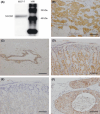
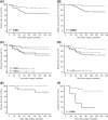
It is well-known that estrogens immensely contribute to the progression of human breast carcinoma, but their detailed molecular mechanisms remain largely unclear. In this study, we identified nucleobindin 2 (NUCB2) as a gene associated with recurrence based on microarray data of estrogen receptor (ER)-positive breast carcinoma cases (n = 10), and subsequent in vitro study showed that NUCB2 expression was upregulated by estradiol in ER-positive MCF-7 cells. However, NUCB2 has not yet been examined in breast carcinoma, and its significance remains unknown. Therefore, we further examined the biological functions of NUCB2 in breast carcinoma using immunohistochemistry and in vitro studies. NUCB2 immunoreactivity was detected in carcinoma cells in 77 of 161 (48%) breast cancer cases, and positively associated with lymph node metastasis and ER status of the patients. In addition, NUCB2 status was significantly associated with an increased risk of recurrence and adverse clinical outcome of the patients using both univariate and multivariate analyses. Results of siRNA transfection experiments showed that NUCB2 significantly increased cell proliferation, and migration and invasion properties in both MCF-7 and ER-negative SK-BR-3 cells. These results suggest that NUCB2 is upregulated by estrogens and plays an important role, especially in the process of metastasis, in breast carcinomas. NUCB2 status is considered a potent prognostic factor in human breast cancer.
© 2011 Japanese Cancer Association.
Figures


Similar articles
-
Nucleobindin 2 (NUCB2) in human endometrial carcinoma: a potent prognostic factor associated with cell proliferation and migration.Endocr J. 2016;63(3):287-99. doi: 10.1507/endocrj.EJ15-0490. Epub 2016 Feb 4. Endocr J. 2016. PMID: 26842712
-
Nudix-type motif 2 in human breast carcinoma: a potent prognostic factor associated with cell proliferation.Int J Cancer. 2011 Apr 15;128(8):1770-82. doi: 10.1002/ijc.25505. Int J Cancer. 2011. PMID: 20533549
-
Nucleobindin 2 expression is an independent prognostic factor for clear cell renal cell carcinoma.Histopathology. 2015 Apr;66(5):650-7. doi: 10.1111/his.12587. Epub 2014 Dec 22. Histopathology. 2015. PMID: 25322808
-
High expression and prognostic role of CAP1 and CtBP2 in breast carcinoma: associated with E-cadherin and cell proliferation.Med Oncol. 2014 Mar;31(3):878. doi: 10.1007/s12032-014-0878-7. Epub 2014 Feb 13. Med Oncol. 2014. PMID: 24522810
-
Om.breast cancer in very young women aged 25 year-old or below in the center of Tunisia and review of the literature.Pathol Oncol Res. 2015 Jul;21(3):553-61. doi: 10.1007/s12253-015-9944-5. Epub 2015 May 12. Pathol Oncol Res. 2015. PMID: 25962349 Review.
Cited by
-
Single Nucleotide Polymorphisms of NUCB2 and their Genetic Associations with Milk Production Traits in Dairy Cows.Genes (Basel). 2019 Jun 13;10(6):449. doi: 10.3390/genes10060449. Genes (Basel). 2019. PMID: 31200542 Free PMC article.
-
Nesfatin-1 is a potential diagnostic biomarker for gastric cancer.Oncol Lett. 2020 Feb;19(2):1577-1583. doi: 10.3892/ol.2019.11200. Epub 2019 Dec 10. Oncol Lett. 2020. PMID: 31966083 Free PMC article.
-
Nucleobindin 2 expression is an independent prognostic factor for bladder cancer.Medicine (Baltimore). 2020 Mar;99(13):e19597. doi: 10.1097/MD.0000000000019597. Medicine (Baltimore). 2020. PMID: 32221080 Free PMC article.
-
Identification of Nucleobindin-2 as a Potential Biomarker for Breast Cancer Metastasis Using iTRAQ-based Quantitative Proteomic Analysis.J Cancer. 2017 Sep 2;8(15):3062-3069. doi: 10.7150/jca.19619. eCollection 2017. J Cancer. 2017. PMID: 28928897 Free PMC article.
-
Association of Irisin/FNDC5 with ERRα and PGC-1α Expression in NSCLC.Int J Mol Sci. 2022 Nov 17;23(22):14204. doi: 10.3390/ijms232214204. Int J Mol Sci. 2022. PMID: 36430689 Free PMC article.
References
-
- Ali S, Coombes RC. Endocrine‐responsive breast cancer and strategies for combating resistance. Nat Rev Cancer 2002; 2: 101–12. - PubMed
-
- Suzuki T, Inoue A, Miki Y et al. Early growth responsive gene 3 in human breast carcinoma: a regulator of estrogen‐meditated invasion and a potent prognostic factor. Endocr Relat Cancer 2007; 14: 279–92. - PubMed
-
- Frasor J, Danes JM, Komm B, Chang KC, Lyttle CR, Katzenellenbogen BS. Profiling of estrogen up‐ and down‐regulated gene expression in human breast cancer cells: insights into gene networks and pathways underlying estrogenic control of proliferation and cell phenotype. Endocrinology 2003; 144: 4562–74. - PubMed
-
- Hayashi SI, Eguchi H, Tanimoto K et al. The expression and function of estrogen receptor alpha and beta in human breast cancer and its clinical application. Endocr Relat Cancer 2003; 10: 193–202. - PubMed
Publication types
MeSH terms
Substances
LinkOut - more resources
Full Text Sources
Medical
Molecular Biology Databases

