

White-tailed deer are susceptible to the agent of sheep scrapie by intracerebral inoculation
- PMID: 21988781
- PMCID: PMC3199251
- DOI: 10.1186/1297-9716-42-107
White-tailed deer are susceptible to the agent of sheep scrapie by intracerebral inoculation
Abstract
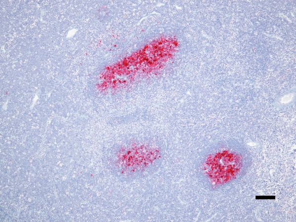
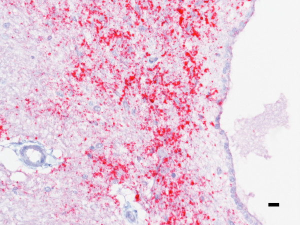
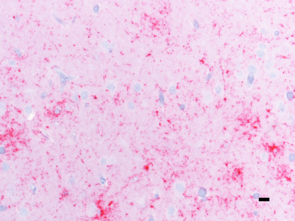
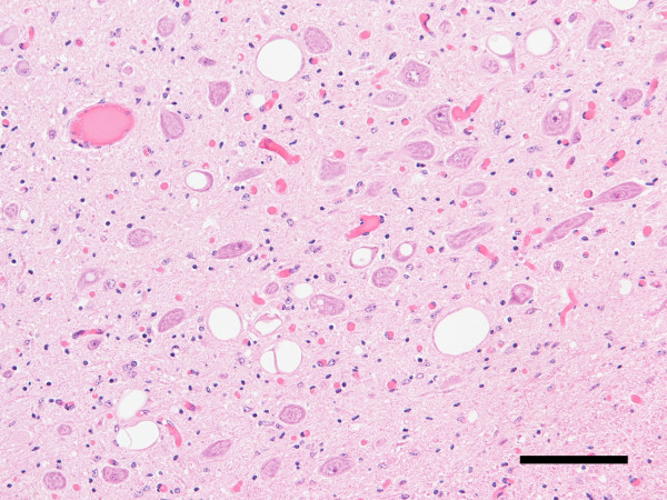
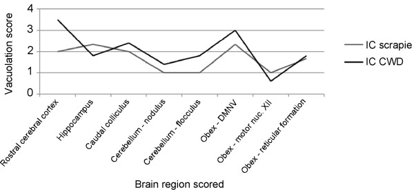
Interspecies transmission studies afford the opportunity to better understand the potential host range and origins of prion diseases. The purpose of this experiment was to determine susceptibility of white-tailed deer to the agent of scrapie after intracerebral inoculation and to compare clinical signs and lesions to those reported for chronic wasting disease (CWD). Deer (n = 5) were inoculated with 1 mL of a 10% (wt/vol) brain homogenate derived from a sheep clinically affected with scrapie. A non-inoculated deer was maintained as a negative control. Deer were observed daily for clinical signs of disease and euthanized and necropsied when unequivocal signs of scrapie were noted. One animal died 7 months post inoculation (pi) due to intercurrent disease. Examinations of brain tissue for the presence of the disease-associated abnormal prion protein (PrP(Sc)) by western blot (WB) and immunohistochemistry (IHC) were negative whereas IHC of lymphoid tissues was positive. Deer necropsied at 15-22 months pi were positive for scrapie by IHC and WB. Deer necropsied after 20 months pi had clinical signs of depression and progressive weight loss. Tissues with PrP(Sc) immunoreactivity included brain (at levels of cerebrum, hippocampus, colliculus, cerebellum, and brainstem), trigeminal ganglion, neurohypophysis, retina, spinal cord, and various lymphoid tissues including tonsil, retropharyngeal and mesenteric lymph nodes, Peyer's patches, and spleen. This work demonstrates for the first time that white-tailed deer are susceptible to sheep scrapie by intracerebral inoculation. To further test the susceptibility of white-tailed deer to scrapie these experiments will be repeated with a more natural route of inoculation.
Figures
Similar articles
-
Raccoons accumulate PrPSc after intracranial inoculation of the agents of chronic wasting disease or transmissible mink encephalopathy but not atypical scrapie.J Vet Diagn Invest. 2019 Mar;31(2):200-209. doi: 10.1177/1040638718825290. Epub 2019 Jan 29. J Vet Diagn Invest. 2019. PMID: 30694116 Free PMC article.
-
Characterization of Classical Sheep Scrapie in White-tailed Deer after Experimental Oronasal Exposure.J Infect Dis. 2023 Jun 15;227(12):1386-1395. doi: 10.1093/infdis/jiac443. J Infect Dis. 2023. PMID: 36344485
-
Detection of PrPSc in lymphoid tissues of lambs experimentally exposed to the scrapie agent.J Comp Pathol. 2003 Feb-Apr;128(2-3):172-81. doi: 10.1053/jcpa.2002.0625. J Comp Pathol. 2003. PMID: 12634095
-
Scrapie and chronic wasting disease.Clin Lab Med. 2003 Mar;23(1):139-59. doi: 10.1016/s0272-2712(02)00040-9. Clin Lab Med. 2003. PMID: 12733429 Review.
-
Chronic wasting disease of cervids.Curr Top Microbiol Immunol. 2004;284:193-214. doi: 10.1007/978-3-662-08441-0_8. Curr Top Microbiol Immunol. 2004. PMID: 15148993 Review.
Cited by
-
Scientific opinion on chronic wasting disease (II).EFSA J. 2018 Jan 17;16(1):e05132. doi: 10.2903/j.efsa.2018.5132. eCollection 2018 Jan. EFSA J. 2018. PMID: 32625679 Free PMC article.
-
Using White-tailed Deer (Odocoileus virginianus) in Infectious Disease Research.J Am Assoc Lab Anim Sci. 2017 Jul 1;56(4):350-360. J Am Assoc Lab Anim Sci. 2017. PMID: 28724483 Free PMC article.
-
Chronic wasting disease (CWD) in cervids.EFSA J. 2017 Jan 18;15(1):e04667. doi: 10.2903/j.efsa.2017.4667. eCollection 2017 Jan. EFSA J. 2017. PMID: 32625260 Free PMC article.
-
Raccoons accumulate PrPSc after intracranial inoculation of the agents of chronic wasting disease or transmissible mink encephalopathy but not atypical scrapie.J Vet Diagn Invest. 2019 Mar;31(2):200-209. doi: 10.1177/1040638718825290. Epub 2019 Jan 29. J Vet Diagn Invest. 2019. PMID: 30694116 Free PMC article.
-
Scrapie in Swine: a Diagnostic Challenge.Food Saf (Tokyo). 2016 Dec 7;4(4):110-114. doi: 10.14252/foodsafetyfscj.2016019. eCollection 2016 Dec. Food Saf (Tokyo). 2016. PMID: 32231914 Free PMC article. Review.
References
-
- Spraker TR, Miller MW, Williams ES, Getzy DM, Adrian WJ, Schoonveld GG, Spowart RA, O'Rourke KI, Miller JM, Merz PA. Spongiform encephalopathy in free-ranging mule deer (Odocoileus hemionus), white-tailed deer (Odocoileus virginianus) and Rocky Mountain elk (Cervus elaphus nelsoni) in northcentral Colorado. J Wild Dis. 1997;33:1–6. - PubMed
-
- Williams ES, Young S. Chronic wasting disease of captive mule deer: a spongiform encephalopathy. J Wild Dis. 1980;16:89–98. - PubMed
-
- Williams ES, Young S. Spongiform encephalopathy of Rocky Mountain elk. J Wild Dis. 1982;18:465–471. - PubMed
MeSH terms
Substances
LinkOut - more resources
Full Text Sources
Research Materials
Miscellaneous
