. 2011 Oct 11:2:496.
doi: 10.1038/ncomms1502.

Electrosensory ampullary organs are derived from lateral line placodes in bony fishes

Affiliations

Electrosensory ampullary organs are derived from lateral line placodes in bony fishes

Melinda S Modrell et al. Nat Commun. .

Abstract

Electroreception is an ancient subdivision of the lateral line sensory system, found in all major vertebrate groups (though lost in frogs, amniotes and most ray-finned fishes). Electroreception is mediated by 'hair cells' in ampullary organs, distributed in fields flanking lines of mechanosensory hair cell-containing neuromasts that detect local water movement. Neuromasts, and afferent neurons for both neuromasts and ampullary organs, develop from lateral line placodes. Although ampullary organs in the axolotl (a representative of the lobe-finned clade of bony fishes) are lateral line placode-derived, non-placodal origins have been proposed for electroreceptors in other taxa. Here we show morphological and molecular data describing lateral line system development in the basal ray-finned fish Polyodon spathula, and present fate-mapping data that conclusively demonstrate a lateral line placode origin for ampullary organs and neuromasts. Together with the axolotl data, this confirms that ampullary organs are ancestrally lateral line placode-derived in bony fishes.


Figures

Figure 1
Figure 1. Lateral line system of juvenile paddlefish (Polyodon spathula)
(a) Simplified vertebrate phylogeny illustrating the distribution of electroreception within the major vertebrate clades and the position of chondrosteans, which includes paddlefishes and sturgeons (blue box). Red bars: loss of electroreception; green bars: teleost groups that independently evolved electroreception. *Hagfishes are not electroreceptive. (b, c) SEM and schematic, respectively, of a juvenile paddlefish showing the distribution of the mechanoreceptive neuromast canals and electroreceptive ampullary organ fields. In b, ampullary organ fields are grouped by different colours based on their innervation patterns (see panel j). (d) Magnification of boxed area in b. Red arrowhead: surface pore of an ampullary organ; arrow: a neuromast recessed in a canal. (e) Magnification of neuromast (arrow) within the preopercular canal and (f) of the neuromast itself showing kinocilia on the apical surface. (g) Lateral view of posterior lateral line trunk neuromasts (arrows). (h) Transverse section through a neuromast. Black arrowhead: kinocilia; asterisks: central sensory cells. (i) Transverse section through an ampullary organ cluster containing two organs (round nuclei: sensory cells in epithelium). (j) Schematic reconstruction of head and anterior trunk lateral line nerves based on Sudan Black-stained specimens. Diagonal hatching: neuromast sensory canal lines. The three pre-otic ganglia and associated nerves innervate distinct ampullary organ fields: GAD innervates the ds, vs, di, vi fields (yellow); GAV innervates the app and ppp fields (turquoise); GOL innervates the dot and vot fields (teal). (k) Schematic of the brain and lateral line ganglia (dotted areas) and associated nerves based on Sudan Black-stained specimens, sectioned material and an osmicated dissection of juvenile cranial nerves. Circles highlight the roots of pre-otic (green) and post-otic (blue) afferents. Scale bars: 1 mm (b, j, k), 200 μm (d), 100 μm (e, g), 10 μm (f), 25 μm (h, i). Abbreviations: AM, anastomosis of otic ramus of anterodorsal lateral line nerve with middle lateral line nerve; an, anterior naris; ao, ampullary organ; app, anterior preopercular ampullary field; AS, anastomosis of otic ramus of anterodorsal lateral line nerve with middle lateral line nerve and supratemporal ramus of posterior lateral line nerve; ba, barbel; BR, buccal ramus of anterodorsal lateral line nerve; dot, dorsal otic ampullary field; di, dorsal infraorbital ampullary field; DR, dorsal root of anterior lateral line nerves; ds, dorsal supraorbital ampullary field; e, eye; GAD, ganglion of anterodorsal lateral line nerve; GAV, ganglion of anteroventral lateral line nerve; GML, ganglion of middle lateral line nerve; GOL, ganglion of otic lateral line nerve; GPL, ganglion of posterior lateral line nerve; GST, ganglion of supratemporal lateral line nerve; HM, hyomandibular trunk (mixed trunk with large component of anteroventral lateral line fibres); HR, hyoid ramus of hyomandibular trunk; io, infraorbital lateral line; m, middle lateral line; MD, external mandibular ramus of hyomandibular trunk; ML, middle lateral line nerve; N, first neuromast ramule of posterior lateral line nerve; nm, neuromast; ol, otic lateral line; OL, otic lateral line nerve; OP, opercular ramus of hyomandibular trunk; PL posterior lateral line nerve; pll, posterior lateral line; pn, posterior naris; pop, preopercular lateral line; ppp, posterior preopercular field; R, ramule to spiracular organ; RAPP, ramule to ampullary organs of anterior preopercular field; RD, ramule to dorsal infraorbital field posterior to eye; RDIR, ramule to dorsal infraorbital field on rostrum; RDOT, ramule to dorsal otic field; RDS, ramule to dorsal supraorbital field; RPL, root of post-otic lateral line nerves; RPPP, ramule to posterior preopercular field; RV, ramule to ventral otic field; RVIB, ramule to ventral infraorbital field on upper jaw; RVIR, ramule to ventral infraorbital field on rostrum; RVS, ramule to ventral supraorbital field; s, spiracle; sc, spiracular chamber; so, supraorbital lateral line; SR, superficial ophthalmic ramus of anterodorsal lateral line nerve; st, supratemporal lateral line; ST, supratemporal lateral line nerve; vi, ventral infraorbital field; vot, ventral otic field; VR, ventral root of anterior lateral line nerves; vs, ventral supraorbital field.
Figure 2
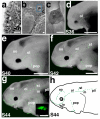
Figure 2. Spatiotemporal development of functional neuromasts
(a) Transverse section through a neuromast primordium developing in the otic lateral line placode at stage 31/32. (b) SEM of a stage 33 embryo showing the first neuromast to erupt (blue box), in the otic lateral line. (c) Magnification of erupted neuromast in b. (d-g) Fluorescent images superimposed on darkfield images of embryos labelled with FM1-43 (green) from stages 36 through 44. Anterior to left. (d) Stage 36 embryo showing the first functional neuromasts in the otic lateral line (ol). (e) Stage 40 embryo showing FM1-43-positive cells in the neuromasts of the supraorbital (so), infraorbital (io) and preopercular (pop) lines. (f) Stage 42 embryo showing neuromasts of the middle (m) and supratemporal (st) lines. (g) Stage 44 embryo. All neuromast lines are present, including the posterior lateral line (pll). Inset: magnification of FM1-43-positive hair cell cluster, showing approximately 10-12 hair cells. (h) Schematic of a stage 44 embryo showing all the neuromast canal lines. Scale bars: 25 μm (a), 10 μm (b, c), 1 mm (d-g). Abbreviations: e, eye; io, infraorbital lateral line; m, middle lateral line; ol; otic lateral line; pll, posterior lateral line; pop, preopercular lateral line; S, stage; so, supraorbital lateral line; st, supratemporal lateral line.
Figure 3
Figure 3. Ampullary organ development
Anterior to left; lateral views unless otherwise noted. (a) Transverse section through an ampullary organ primordium at stage 35. (b) SEM of a stage 39 embryo. Ampullary organs have erupted through the surface in several fields, in particular on the opercular flap. The red arrowheads in panels b-f indicate approximately the same region of ampullary organs that are erupting or have erupted at the different stages of development. The arrows indicate neuromast canal lines, specifically of the otic and preopercular lines. (c) SEM showing a surface view through the pore of a newly erupted ampullary organ. (d) SEM of a stage 42 embryo. Ampullary organs appear to have erupted from all fields by this stage. (e) SEM of a stage 44 embryo showing further differentiation of the ampullary organ fields. (f) Fluorescent image of a stage 46 embryo skin mount, stained with the nuclear marker Sytox Green. Individual clusters of ampullary organs are discernable, as are the neuromast canal lines. (g,h) Confocal images of ampullary organs stained with the nuclear marker DAPI in (g) surface view and (h) transverse view. The F-actin marker phalloidin (green) labels the microvilli-enriched apical surface. (i) Confocal image of a neuromast in transverse view at the same stage for comparison with h. In h and i, the round nuclei of sensory cells are outlined. (j) Stage 42 embryo stained for parvalbumin-3 (Pv3). Hair cells within both neuromasts and ampullary organs express this Ca2+-binding protein. (k) Stage 46 embryo stained for parvalbumin-3. Pv3 is strongly expressed in all lateral line sensory organs. (l, m) False colour overlay with DAPI images, respectively, of transverse sections through ampullary organs and a neuromast. Pv-3 (red in l, m) specifically labels the sensory hair cells in both ampullary organs and neuromasts. Scale bars: 25 μm (a), 100 μm (b), 10 μm (c, l, m), 500 μm (d, e, j, k), 30 μm (h, i). Abbreviations: ao, ampullary organ; e, eye; S, stage; tb, taste bud.
Figure 4
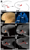
Figure 4. Expression of Eya and Six family members during lateral line development
(a) SEM of a stage 32 embryo. Placodes (outlined) are visible surface bulges. (b) Schematic summarizing histological serial reconstructions at stage 32 of the three pre-otic (anterodorsal [yellow], anteroventral [turquoise], otic [teal]) and three post-otic lateral line placodes (middle, supratemporal, posterior [green]). (c, d) Transverse sections through the anterodorsal placode at the level of the eye (c) and otic vesicle (d). Blue arrowheads indicate extent of thickened epithelium (i.e., the placode). Red arrowhead indicates a developing neuromast primordium. (e-i) Whole-mount in situ hybridisations for Eya1, Eya4, Six1 and Six4 from stages 32-46; schematics show the general expression pattern at those stages. Lateral views; anterior is left. (e) At stage 32, Eya1, Six1 and Six4 are expressed in multiple placodes including olfactory and otic (purple), epibranchial (pink), and lateral line (dark purple); strong expression of Eya4 is restricted to lateral line placodes and the otic vesicle. By this stage, the lateral line placodes have started to elongate or spread along the head. (f) At stage 36, all genes are expressed in developing neuromast canal lines and presumptive ampullary organ fields flanking those lines. (g) At stage 39, expression of all genes continues in elongating lateral line primordia and neuromast canals. Individual ampullary organ fields start to be identifiable. At (h) stage 42 and (i) stage 46, expression of all genes continues in developing lateral line organs as well as in the migrating posterior lateral line primordium. (j) Transverse sections showing Eya4 expression in the developing anterior ventral placode (avp) from stages 32-46. By stage 46, the hair cell marker Pv3 (grey-scale image) overlaps with Eya4 as shown in the false-colour overlay of Eya4 (green) and Pv3 (pink). Scale bars: 100 μm (a, c, d, j), 200 μm (e-i). Abbreviations: adp, anterodorsal placode; avp, anteroventral placode; e, eye; epi, epibranchial placode region; hb, hindbrain; mb, midbrain; mlp, middle lateral line placode; olf, olfactory; otp, otic lateral line placode; ov, otic vesicle; plp, posterior lateral line placode; S, stage; stp, supratemporal placode.
Figure 5
Figure 5. In vivo labelling of lateral line placodes
(a) Schematic of in vivo labelling experimental design. Embryos are focally injected with DiI between stage 32 and 34 and followed to stage 46. (b) Stage 32 embryo immediately after focal injection with DiI. In this example, the anterodorsal placode (outlined in red) was labelled. (c) Transverse section through another stage 32 embryo immediately after injection, showing DiI-positive cells (red; DAPI in blue) in the anterodorsal placode. (d-g) Fluorescent images superimposed on darkfield images of the same embryo shown in panel b, at successive stages of development. (d) Stage 36. (e) Stage 39. (f) Stage 42. (g) Stage 46. As development progresses, DiI spreads anteriorly from the site of injection as neuromasts of the infraorbital line (arrow) are deposited. Ampullary organs (red arrowhead) are also labelled near the site of injection. Scale bars: 500 μm (b), 100 μm (c), 1 mm (d-g). Abbreviations: adp, anterodorsal lateral line placode; e, eye; hb, hindbrain; olf, olfactory; ov, otic vesicle; S, stage.
Figure 6
Figure 6. Lateral line placodes give rise to ampullary organs and neuromasts
(a) Stage 32 embryo after focal injection with DiI in the anterodorsal placode; injection site outlined in red. (b) Same embryo at stage 46, showing DiI-labelled cells in the infraorbital neuromasts and dorsal infraorbital and ventral infraorbital ampullary organ fields. (c, d) Transverse sections through (c) a DiI (red)-labelled neuromast and (d) DiI-labelled ampullary organs, counterstained with Sytox Green. (e) Stage 32 embryo after focal injection with DiI in the anteroventral placode; injection site outlined in red. (f) Same embryo at stage 46, showing DiI-labelled cells in preopercular neuromasts and flanking ampullary organs. (g) Opercular skin mount from embryo in (f), counterstained with Sytox Green, showing DiI in preopercular neuromasts and flanking ampullary organs. (h) Magnification of DiI-labelled neuromasts from preopercular neuromast line in (g). (i) Magnification of DiI-labelled ampullary organ from posterior preopercular field in (g). The plane of focus is the sensory cell epithelium. (j) Stage 34 embryo after focal injection with DiI in the anteroventral placode; injection site outlined in red. (k) Same embryo at stage 46, showing DiI-labelled cells in a subset of preopercular neuromasts and flanking ampullary organs. (l) Confocal image of transverse section from embryo in (k), stained with phalloidin (green) and DAPI (blue), showing a DiI (red)-labelled ampullary organ. (m) Magnification from (l), showing DiI (red) within the sensory epithelium. (n) Stage 32 embryo after focal injection with DiI in the anterodorsal placode; injection site outlined in red. (o) Same embryo at stage 46, showing DiI-labelled cells in infraorbital line neuromasts and flanking ampullary organs. (p) Transverse section through head of embryo in (o), stained with Sytox Green. (q) Magnification from (p) showing DiI-labelled cells (red) in the anterodorsal lateral line ganglion. Scale bars: 500 μm (a, e, j, n), 1 mm (b, f, k, o), 50 μm (c, d, h, i, q), 100 μm (g), 30 μm (l), 250 μm (p). Abbreviations: ao, ampullary organ; e, eye; GAD, anterodorsal lateral line ganglion; hb, hindbrain; io, infraorbital lateral line; nm, neuromast; ov, otic vesicle; p, pharynx; pop, preopercular lateral line.

Similar articles

Cited by

References

    1. Coombs S, Montgomery JC. The enigmatic lateral line system. In: Popper AN, Fay RR, editors. Comparative Hearing: Fishes and Amphibians. Springer-Verlag; New York: 1999. pp. 319–362.
    1. Bullock TH, Hopkins CD, Popper AN, Fay RR. Electroreception. Springer; New York: 2005.
    1. Bodznick D, Montgomery JC. The physiology of low-frequency electrosensory systems. In: Bullock TH, Hopkins CD, Popper AN, Fay RR, editors. Electroreception. Springer; New York: 2005. pp. 132–153.
    1. Jørgensen JM. Morphology of electroreceptive sensory organs. In: Bullock TH, Hopkins CD, Popper AN, Fay RR, editors. Electroreception. Springer; New York: 2005. pp. 47–67.
    1. Northcutt RG. Evolution of gnathostome lateral line ontogenies. Brain Behav. Evol. 1997;50:25–37. - PubMed