. 2012 May;1821(5):727-35.
doi: 10.1016/j.bbalip.2011.09.013. Epub 2011 Oct 2.

Gut triglyceride production

Affiliations

Gut triglyceride production

Xiaoyue Pan et al. Biochim Biophys Acta. 2012 May.

Abstract

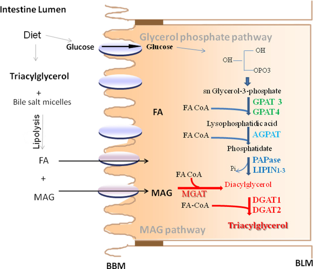
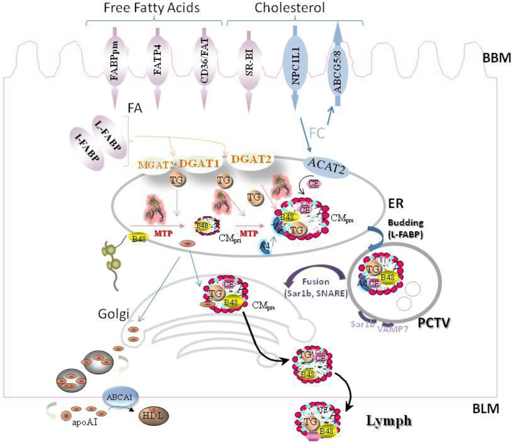
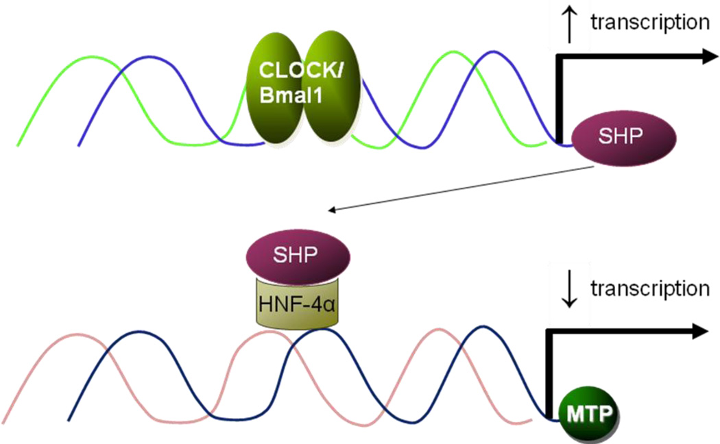
Our knowledge of how the body absorbs triacylglycerols (TAG) from the diet and how this process is regulated has increased at a rapid rate in recent years. Dietary TAG are hydrolyzed in the intestinal lumen to free fatty acids (FFA) and monoacylglycerols (MAG), which are taken up by enterocytes from their apical side, transported to the endoplasmic reticulum (ER) and resynthesized into TAG. TAG are assembled into chylomicrons (CM) in the ER, transported to the Golgi via pre-chylomicron transport vesicles and secreted towards the basolateral side. In this review, we mainly focus on the roles of key proteins involved in uptake and intracellular transport of fatty acids, their conversion to TAG and packaging into CM. We will also discuss intracellular transport and secretion of CM. Moreover, we will bring to light few factors that regulate gut triglyceride production. Furthermore, we briefly summarize pathways involved in cholesterol absorption. This article is part of a Special Issue entitled Triglyceride Metabolism and Disease.


Figures

Fig 1
Fig 1. Biosynthesis of triacylglycerols in enterocytes
Dietary TAG are solubilized in bile salt micelles and lipolysed in the intestinal lumen to free fatty acid and monoacylglycerol (MAG) that are taken up by enterocytes involving diffusion and protein meditated uptake, transported to the ER by carrier proteins and used for the synthesis of TAG by MAG pathway (red line, red). In this pathway, MAG and free fatty acids are first converted to DAG by monoacylglycerol:acylCoA acyltransferase (MGAT) enzymes. DAG is then converted to TAG by diacylglyerol:acylcoA acyltarnsfrease 1 and 2 (DGAT1 and DGAT2). TAG are also synthesized from glucose by the glycerol-3-phosphate pathway. In this pathway, GPAT3/GPAT4 catalyze the esterification of glycerol-3-phosphate to form lysophosphatidic acid. 1-acyl-sn-glycerol-3-phosphate acyltransferases (AGPAT1-5; also called LPAAT) esterify lysophosphatidic acid to form phosphatidate. Three isoforms of phosphatidic acid phosphohydrolase (PAPase, also known as lipins 1–3), hydrolyze the phosphate to form diacylglycerol (DAG). DAG is then converted to TAG by DGATs as in the MAG pathway.
Fig 2
Fig 2. Cellular mechanisms involved in intestinal TAG absorption
Apart from diffusion, the transport of free fatty acids (FA) across the brush border membrane (BBM) is facilitated by different proteins, such as plasma membrane fatty acid binding protein (FABPpm), fatty acid transport protein 4 (FATP4), and fatty acid translocase (FAT/CD36). It is then translocated to the endoplasmic reticulum (ER) by cytosolic fatty acid binding proteins (L-FABP and I-FABP). MGAT and DGAT enzymes then convert MAG and FA to TAG with an intermediate synthesis of DAG. Cholesterol uptake is facilitated by Niemann-Pick C-1-like 1 (NPC1L1). There is evidence for the involvement of other proteins in this pathway such as SR-B1. In the intracellular compartment, free cholesterol has two fates. It can be exported back to the intestinal lumen by ATP-binding cassette transporters G5 and G8 (ABCG5/8) or converted to cholesterol esters by acyl CoA cholesterol acyltransferases 1& 2 (ACAT1 & ACAT2). Newly translated apolipoprotein B (apoB-48) is lipidated by microsomal triglyceride transport protein (MTP) to form primordial chylomicrons (CM) that is further expanded in size by the addition of TAG in the core by MTP to form a pre-CM. These particles also acquire apoAIV at this time. Prechylomicrons are concentrated and exported from the ER in PCTVs. L-FABP facilitates the budding of PCTV from the ER. These vesicles then fuse with the cis-Golgi in a process that requires VAMP7. In the Golgi, apoA1 is added to prechylomicrons. Later, these particles are transported to plasma membrane using different transport vesicles and are ultimately released towards the basolateral side by enterocytes. TAG are exclusively transported via the CM pathway. By contrast, cholesterol is transported across the intestinal epithelial cells by two pathways. One pathway is the same as the CM pathway. In this pathway free cholesterol and esterified cholesterol are added onto CM by MTP. In the second apoB-independent pathway, cholesterol is secreted involving ABCA1 and apoAI. ApoAI is synthesized in the ER and transported independent of CMs to the Golgi where some of it associates with CMs and the rest is secreted independent of CMs. The free apoAI might play a role in cholesterol transport.
Fig 3
Fig 3. Regulation of MTP by Clock via SHP
Clock represses MTP expression through an indirect mechanism. Clock/Bmal1 binds to the E-Box in the small heterodimer partner (SHP) promoter and increases its expression in the daytime. SHP, in turns, interacts with transcription factors/enhancer present on the MTP promoter and represses its activity leading to decreases in day time.

Similar articles

Cited by

References

    1. Abumrad NA, el-Maghrabi MR, Amri EZ, Lopez E, Grimaldi PA. Cloning of a rat adipocyte membrane protein implicated in binding or transport of long-chain fatty acids that is induced during preadipocyte differentiation. Homology with human CD36. J. Biol. Chem. 1993;268:17665. - PubMed
    1. Adeli K, Lewis GF. Intestinal lipoprotein overproduction in insulin-resistant states. Curr. Opin. Lipidol. 2008;19:221. - PubMed
    1. Agellon LB, Drozdowski L, Li L, Iordache C, Luong L, Clandinin MT, Uwiera RR, Toth MJ, Thomson AB. Loss of intestinal fatty acid binding protein increases the susceptibility of male mice to high fat diet-induced fatty liver. Biochim. Biophys. Acta. 2007;1771:1283. - PubMed
    1. Alpers DH, Bass NM, Engle MJ, Schryver-Kecskemeti K. Intestinal fatty acid binding protein may favor differential apical fatty acid binding in the intestine. Biochim. Biophys. Acta. 2000;1483:352. - PubMed
    1. Alpers DH, Strauss AW, Ockner RK, Bass NM, Gordon JI. Cloning of a cDNA encoding rat intestinal fatty acid binding protein. Proc. Natl. Acad. Sci. U. S. A. 1984;81:313. - PMC - PubMed

Publication types