Comparative Study
. 2011 Oct 11:6:33.
doi: 10.1186/1749-8104-6-33.

Type I vs type II spiral ganglion neurons exhibit differential survival and neuritogenesis during cochlear development

Affiliations
Comparative Study

Type I vs type II spiral ganglion neurons exhibit differential survival and neuritogenesis during cochlear development

Meagan Barclay et al. Neural Dev. .

Abstract

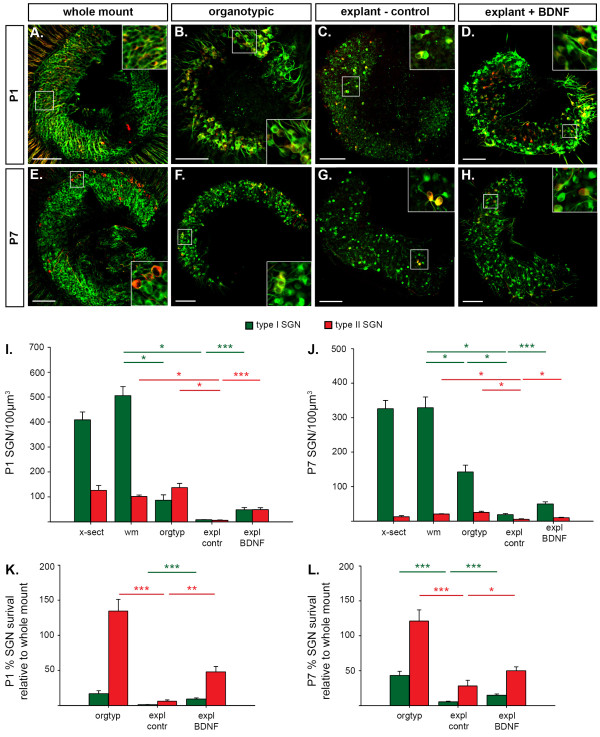
Background: The mechanisms that consolidate neural circuitry are a major focus of neuroscience. In the mammalian cochlea, the refinement of spiral ganglion neuron (SGN) innervation to the inner hair cells (by type I SGNs) and the outer hair cells (by type II SGNs) is accompanied by a 25% loss of SGNs.

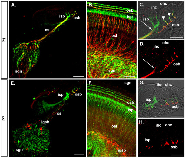
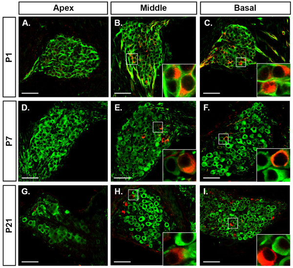
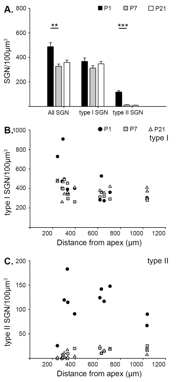
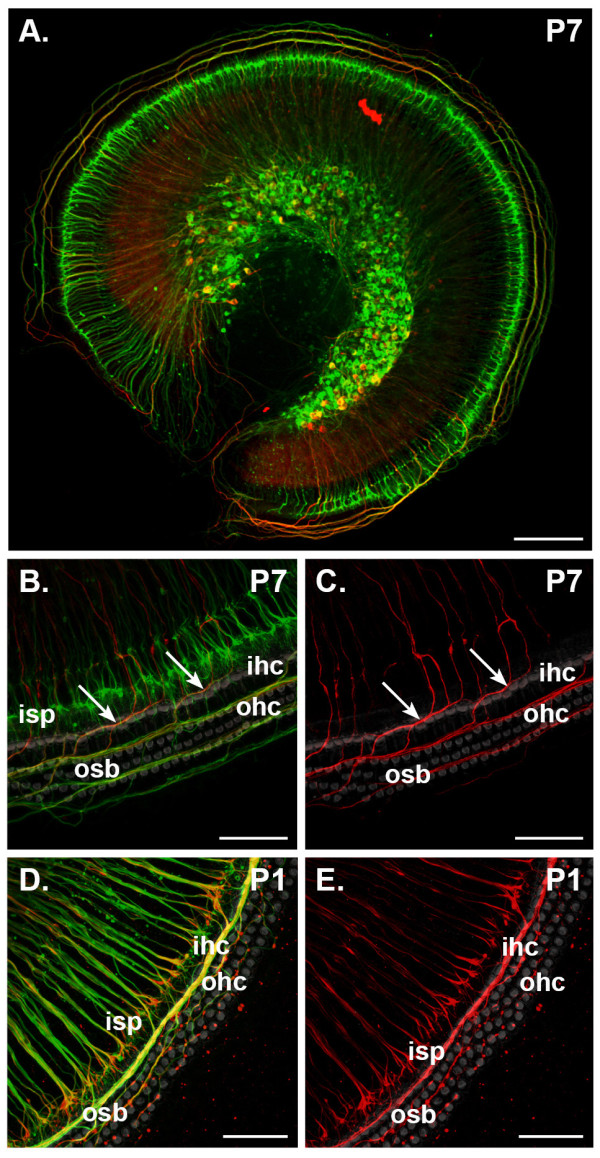
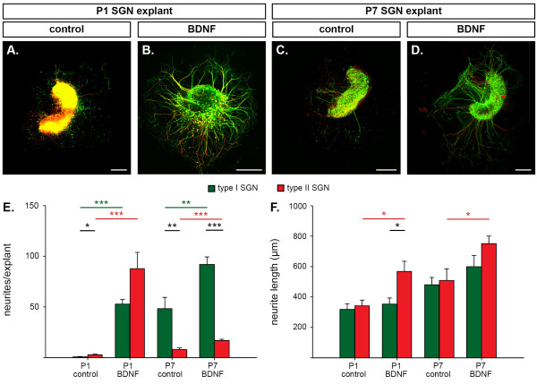
Results: We investigated the segregation of neuronal loss in the mouse cochlea using β-tubulin and peripherin antisera to immunolabel all SGNs and selectively type II SGNs, respectively, and discovered that it is the type II SGN population that is predominately lost within the first postnatal week. Developmental neuronal loss has been attributed to the decline in neurotrophin expression by the target hair cells during this period, so we next examined survival of SGN sub-populations using tissue culture of the mid apex-mid turn region of neonatal mouse cochleae. In organotypic culture for 48 hours from postnatal day 1, endogenous trophic support from the organ of Corti proved sufficient to maintain all type II SGNs; however, a large proportion of type I SGNs were lost. Culture of the spiral ganglion as an explant, with removal of the organ of Corti, led to loss of the majority of both SGN sub-types. Brain-derived neurotrophic factor (BDNF) added as a supplement to the media rescued a significant proportion of the SGNs, particularly the type II SGNs, which also showed increased neuritogenesis. The known decline in BDNF production by the rodent sensory epithelium after birth is therefore a likely mediator of type II neuron apoptosis.

Conclusion: Our study thus indicates that BDNF supply from the organ of Corti supports consolidation of type II innervation in the neonatal mouse cochlea. In contrast, type I SGNs likely rely on additional sources for trophic support.


Figures

Figure 1
Figure 1
Double labeling for β-tubulin and peripherin distinguishes type I and type II spiral ganglion neurons (SGNs) and their peripheral innervation during the first postnatal week of mouse development. Single plane (A,C,D,E,G,H) and maximum projections (B,F) of confocal image stacks show β-tubulin (green) and peripherin (red) immunofluorescence in cochleae from P1 (A-D) and P7 (E-H) mice. (A-D) At P1 peripheral fibers from type I SGNs (immunolabeled for β-tubulin only) form the inner spiral plexus (isp) beneath the inner hair cell (ihc). Type II SGN soma and peripheral fibers (β-tubulin and peripherin immunolabeled) are numerous in the spiral ganglion (sgn) and osseus spiral lamina (osl) of P1 cochleae (A,B). Their neurites are present beneath the IHCs (arrow in (C,D)) and beneath the outer hair cells (ohc) in the outer spiral bundles (osb). (E-H) At P7, the majority of neurons in the spiral ganglion are type I neurons (E,F) and their neurites innervate the IHCs (G). Peripherin and β-tubulin double-immunolabeled type II SGNs are now localized laterally in the spiral ganglion near the intra-ganglionic spiral bundle (igsb), their fibers form the outer spiral bundle and innervate the OHC only; no peripherin-immunolabeled fibers extend toward the IHC (H). Variation in the color (yellow to red) of double labeled (type II) neuron soma and their neurite processes arises from differences in the relative intensity of the β-tubulin (green fluorescence) in soma and neurites and also peripherin (red) fluorescence in merged confocal images. This is particularly evident at P7, where the type II soma appear red as there is a decline in β-tubulin labeling, whereas the outer spiral bundle region is yellow, due to approximate equivalence of the intensity of the red and green immunofluorescence in this region (see (E)). We attribute double labeling (type II SGNs) versus single (β-tubulin) labeling (type I SGNs) from analysis of individual channels. Panels (A,C-E,G,H) are from cryosectioned tissue; (B,F) are whole-mount preparations. Scale bars: 50 μm (A,B,E,F); 25 μm (C,D,G,H).
Figure 2
Figure 2
Peripherin immunolabelled type II spiral ganglion neurons (SGNs) are absent at the apex of the cochlea and between P1 and P7 their number reduces in the middle and basal regions. (A-I) Single plane confocal images of β-tubulin (green) and peripherin (red) double immunolabeling of SGNs in the most apical region (apex) (A,D,G), and in the middle (B,E,H) and basal (C,F,I) turns in sectioned cochleae from P1 (A-C), P7 (D-E) and P21 (G-I) mice. Marked areas are shown in insets and detail β-tubulin and peripherin immunolabeling of SGNs. β-Tubulin and peripherin double immunolabeled type II SGNs are absent from the apex of P1 (A), P7 (D) and P21 (G) mouse cochleae. Many of these double-immunolabeled type II SGNs are present in the middle and basal turns in P1 cochlea (B,C); however, this number is reduced in P7 (E,F) and P21 (H,I) cochleae. The level of β-tubulin expression in the type II SGN soma appears reduced in P7 and P21 cochleae, resulting in relatively stronger red signal (peripherin channel). Scale bars: 50 μm.
Figure 3
Figure 3
Quantitative analysis of type I and type II spiral ganglion neuron (SGN) densities in P1, P7 and P21 mouse cochlea shows selective loss of type II SGNs in the first postnatal week in vivo. (A) Density of all SGNs, type I SGNs and type II SGNs calculated from cell counts taken from mid-modiolar cross-sections from P1 (black), P7 (grey) and P21 (white) mouse cochlea. Mean density ± standard error of the mean; ANOVA, post hoc Holm-Sidak test, **P < 0.01, ***P < 0.001. (B,C) The density of type I (B) and type II (C) SGNs within different regions of the spiral ganglion in mid-modiolar cross-sections are plotted against the distance of the ganglia from the apex of the cochlea.
Figure 4
Figure 4
The expression pattern of β-tubulin and peripherin in type I and type II spiral ganglion neurons is maintained in vitro. Maximum intensity projections from confocal z-stacks show immunofluorescence labeling for β-tubulin (green) and peripherin (red) in the mid-apex-mid-turn region of P1 (D,E) and P7 (A-C) mouse cochlea following 48 hour organotypic culture (SGNs and organ of Corti intact). Rhodamine-phalloidin labeling (grey in (B-E)) confirmed the survival of target hair cells. (A,B) Immunolabeling of organotypic cultures of P7 tissue shows the inner spiral plexus (isp), formed by type I fibers, contains β-tubulin protein only (detailed in (B)), whereas the outer spiral bundles (osb) that arise from type II fibers express both β-tubulin and peripherin (arrows in (B,C)). (C) Examination of peripherin immunofluorescence alone confirms that this protein is expressed only in fibers that cross the tunnel of Corti and innervate the outer hair cells (ohc). (D,E) Confocal imaging of organotypic culture of P1 cochlear tissue shows that singularly β-tubulin immunofluorescent fibers beneath the inner hair cells (ihc) are reduced in density compared to the in vivo situation (compare (D) with Figure 1B). A large portion of these fibers co-immunolabel for peripherin (D,E). The outer spiral bundle double immunolabels with β-tubulin and peripherin (D,E). Scale bars: 150 μm (A); 50 μm (C-E).
Figure 5
Figure 5
The organ of Corti (oC) maintains type II spiral ganglion neurons (SGNs), but not type I SGNs in organotypic culture and brain-derived neurotrophic factor (BDNF) rescues a greater proportion of type II SGNs following explant culture without the oC. (A- H) Single plane confocal images of β-tubulin (green) and peripherin (red) immunofluorescence in the mid-apex-mid-turn region of cochleae from P1 (A-D) and P7 (E-H) mice enabled analysis of type I SGN (immunolabel for β-tubulin only) and type II SGN (immunolabel for β-tubulin and peripherin) density. SGN density in the mid-apex-mid-turn region in vivo was determined in whole mount tissue (whole mount) (A,E). Survival of SGN sub-types was subsequently established following 48 hour culture of cochlear tissue: retaining both SGNs and oC (organotypic) (B,F); just the spiral ganglion, following removal of the oC (explant - control) (C,G) and this spiral ganglion explant cultured with 100 ng/ml BDNF (explant + BDNF) (D,H). Inset shows marked area in corresponding panel at higher magnification and shows detail of type I and type II SGNs. (I,J) Neuron densities for type I (green) and type II (red) SGNs from P1 (I) and P7 (J) mice are plotted for each condition shown in panels (A-H). x-sect, mid-apex-mid-turn ganglion in mid-modiolar cross-sections; wm, whole mount; orgtyp, organotypic; expl BDNF, explant + BDNF; expl contr, explant - control. Mean density and standard error of the mean; one-way ANOVA with post hoc Holm Sidak tests compare SGN survival in the wm, orgtyp and expl contr; unpaired t-tests compare expl contr and expl BDNF; *P < 0.05, ***P < 0.001. (K,L) Percentage survival of type I and type II SGNs in orgtyp, expl BDNF and expl contr relative to mean in vivo whole mount densities are plotted for P1 (K) and P7 (L) mice. Mean density and standard error of the mean; unpaired t-test, *P < 0.05, **P < 0.01, ***P < 0.001. Scale bars: 250 μm.
Figure 6
Figure 6
Brain-derived neurotrophic factor (BDNF) promotes neuritogenesis of both type I and type II spiral ganglion neurons (SGNs) in explants, but has a greater effect on type II SGNs. (A-D) Analysis of neurite outgrowth in mid-apical-mid-turn region explants from P1 (A,B) and P7 (C,D) mouse cochlea following 48 hour culture in control culture media (A,C) or with 100 ng/ml BDNF added to the media (B,D). Confocal immunofluorescence images show type I SGN neurites (β-tubulin (green) only) and type II SGN neurites (β-tubulin and peripherin (red)). (E,F) BDNF stimulated an increased number of type I (green) and type II (red) neurites per explant (E) and significantly longer type II neurites (F). The neurites extend from the explants in bundles and then separate into individual fibers. This gives the appearance of branching towards the ends in the low magnification images. Data are represented as mean and standard error of the mean; unpaired t-test, *P < 0.05, **P < 0.01, ***P < 0.001. Scale bars: 250 μm.

References

    1. Kano M, Hashimoto K. Synapse elimination in the central nervous system. Curr Opin Neurobiol. 2009;19:154–161. doi: 10.1016/j.conb.2009.05.002. - DOI - PubMed
    1. Luo L, O'Leary DD. Axon retraction and degeneration in development and disease. Annu Rev Neurosci. 2005;28:127–156. doi: 10.1146/annurev.neuro.28.061604.135632. - DOI - PubMed
    1. Buss RR, Sun W, Oppenheim RW. Adaptive roles of programmed cell death during nervous system development. Annu Rev Neurosci. 2006;29:1–35. doi: 10.1146/annurev.neuro.29.051605.112800. - DOI - PubMed
    1. Clarke PG, Posada A, Primi MP, Castagne V. Neuronal death in the central nervous system during development. Biomed Pharmacother. 1998;52:356–362. doi: 10.1016/S0753-3322(99)80002-X. - DOI - PubMed
    1. Jagger DJ, Housley GD. Membrane properties of type II spiral ganglion neurones identified in a neonatal rat cochlear slice. J Physiol. 2003;552:525–533. doi: 10.1111/j.1469-7793.2003.00525.x. - DOI - PMC - PubMed

Publication types

MeSH terms

Substances