

Differences between the neurogenic and proliferative abilities of Müller glia with stem cell characteristics and the ciliary epithelium from the adult human eye
- PMID: 21989110
- PMCID: PMC3268355
- DOI: 10.1016/j.exer.2011.09.015
Differences between the neurogenic and proliferative abilities of Müller glia with stem cell characteristics and the ciliary epithelium from the adult human eye
Abstract
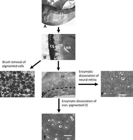
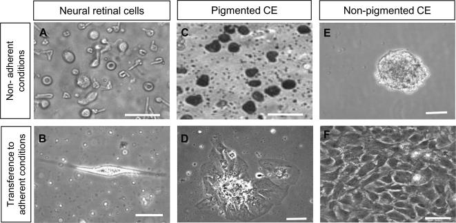
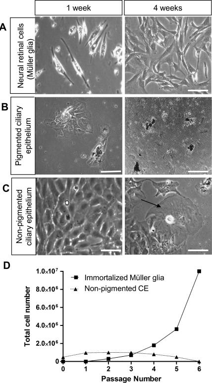
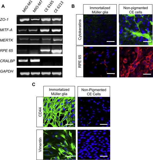
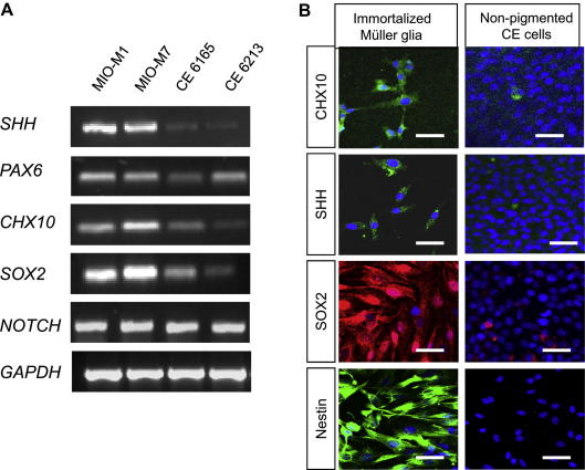
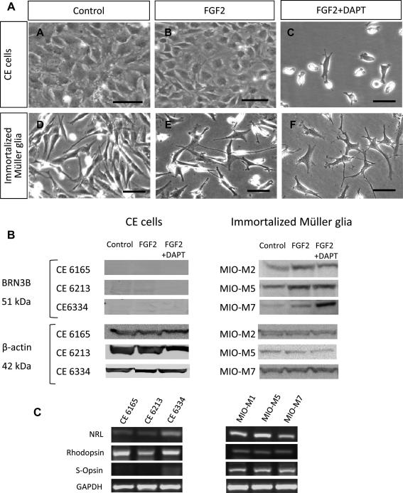
Much controversy has arisen on the nature and sources of stem cells in the adult human retina. Whilst ciliary epithelium has been thought to constitute a source of neural stem cells, a population of Müller glia in the neural retina has also been shown to exhibit neurogenic characteristics. This study aimed to compare the neurogenic and proliferative abilities between these two major cell populations. It also examined whether differences exist between the pigmented and non-pigmented ciliary epithelium (CE) from the adult human eye. On this basis, Müller glia with stem cell characteristics and pigmented and non-pigmented CE were isolated from human neural retina and ciliary epithelium respectively. Expression of glial, epithelial and neural progenitor markers was examined in these cells following culture under adherent and non-adherent conditions and treatments to induce neural differentiation. Unlike pigmented CE which did not proliferate, non-pigmented CE cells exhibited limited proliferation in vitro, unless epidermal growth factor (EGF) was present in the culture medium to prolong their survival. In contrast, Müller glial stem cells (MSC) cultured as adherent monolayers reached confluence within a few weeks and continued to proliferative indefinitely in the absence of EGF. Both MSC and non-pigmented CE expressed markers of neural progenitors, including SOX2, PAX6, CHX10 and NOTCH. Nestin, a neural stem cell marker, was only expressed by MSC. Non-pigmented CE displayed epithelial morphology, limited photoreceptor gene expression and stained strongly for pigmented epithelial markers upon culture with neural differentiation factors. In contrast, MSC adopted neural morphology and expressed markers of retinal ganglion cells and photoreceptors when cultured under similar conditions. This study provides the first demonstration that pigmented CE possess different proliferative abilities from non-pigmented CE. It also showed that although non-pigmented CE express genes of retinal progenitors, they do not differentiate into neurons in vitro, as that seen with Müller glia that proliferate indefinitely in vitro and that acquire markers of retinal neurons in culture under neural differentiation protocols. From these observations it is possible to suggest that Müller glia that express markers of neural progenitors and become spontaneously immortalized in vitro constitute a potential source of retinal neurons for transplantation studies and fulfil the characteristics of true stem cells due to their proliferative and neurogenic ability.
Copyright © 2011 Elsevier Ltd. All rights reserved.
Figures
Similar articles
-
Distribution of Müller stem cells within the neural retina: evidence for the existence of a ciliary margin-like zone in the adult human eye.Exp Eye Res. 2009 Sep;89(3):373-82. doi: 10.1016/j.exer.2009.04.005. Epub 2009 Apr 18. Exp Eye Res. 2009. PMID: 19379736
-
Activation of neural progenitor cells in human eyes with proliferative vitreoretinopathy.Exp Eye Res. 2012 May;98:28-36. doi: 10.1016/j.exer.2012.03.008. Epub 2012 Mar 21. Exp Eye Res. 2012. PMID: 22465407
-
Proliferative Cells Isolated from the Adult Human Peripheral Retina only Transiently Upregulate Key Retinal Markers upon Induced Differentiation.Curr Eye Res. 2018 Mar;43(3):340-349. doi: 10.1080/02713683.2017.1403630. Epub 2017 Nov 21. Curr Eye Res. 2018. PMID: 29161152
-
Müller glia: Stem cells for generation and regeneration of retinal neurons in teleost fish.Prog Retin Eye Res. 2014 May;40:94-123. doi: 10.1016/j.preteyeres.2013.12.007. Epub 2014 Jan 8. Prog Retin Eye Res. 2014. PMID: 24412518 Free PMC article. Review.
-
Retinal regeneration in birds and mice.Curr Opin Genet Dev. 2016 Oct;40:57-64. doi: 10.1016/j.gde.2016.05.028. Epub 2016 Jul 2. Curr Opin Genet Dev. 2016. PMID: 27379897 Review.
Cited by
-
Rekindling Vision: Innovative Strategies for Treating Retinal Degeneration.Int J Mol Sci. 2025 Apr 25;26(9):4078. doi: 10.3390/ijms26094078. Int J Mol Sci. 2025. PMID: 40362317 Free PMC article. Review.
-
GABA maintains the proliferation of progenitors in the developing chick ciliary marginal zone and non-pigmented ciliary epithelium.PLoS One. 2012;7(5):e36874. doi: 10.1371/journal.pone.0036874. Epub 2012 May 9. PLoS One. 2012. PMID: 22590629 Free PMC article.
-
Regeneration of cone photoreceptors when cell ablation is primarily restricted to a particular cone subtype.PLoS One. 2013;8(1):e55410. doi: 10.1371/journal.pone.0055410. Epub 2013 Jan 30. PLoS One. 2013. PMID: 23383182 Free PMC article.
-
Adult Stem Cells, Tools for Repairing the Retina.Curr Ophthalmol Rep. 2019 Mar;7(1):21-29. doi: 10.1007/s40135-019-00195-z. Epub 2019 Jan 24. Curr Ophthalmol Rep. 2019. PMID: 31667009 Free PMC article.
-
Fate bias during neural regeneration adjusts dynamically without recapitulating developmental fate progression.Neural Dev. 2017 Jul 13;12(1):12. doi: 10.1186/s13064-017-0089-y. Neural Dev. 2017. PMID: 28705258 Free PMC article.
References
-
- Ahmad I., Tang L., Pham H. Identification of neural progenitors in the adult mammalian eye. Biochem. Biophys. Res. Commun. 2000;270:517–521. - PubMed
-
- Bertazolli-Filho R., Ghosh S., Huang W., Wollmann G., Coca-Prados M. Molecular evidence that human ocular ciliary epithelium expresses components involved in phototransduction. Biochem. Biophys. Res. Commun. 2001;284:317–325. - PubMed
-
- Das A.V., James J., Rahnenfuhrer J., Thoreson W.B., Bhattacharya S., Zhao X., Ahmad I. Retinal properties and potential of the adult mammalian ciliary epithelium stem cells. Vis. Res. 2005;45:1653–1666. - PubMed
Publication types
MeSH terms
Substances
Grants and funding
LinkOut - more resources
Full Text Sources
Other Literature Sources
Medical
