

The dynamic structure of arterioles
- PMID: 21989114
- PMCID: PMC4435689
- DOI: 10.1111/j.1742-7843.2011.00813.x
The dynamic structure of arterioles
Abstract
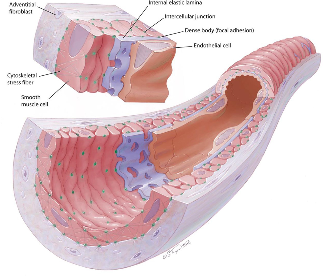
Arterioles are the blood vessels in the arterial side of the vascular tree that are located proximal to the capillaries and, in conjunction with the terminal arteries, provide the majority of resistance to blood flow. Consequently, arterioles are important contributors to the regulation of mean arterial pressure and tissue perfusion. Their wall consists of cellular and extracellular components that have been traditionally classified as conforming three layers: an intima containing endothelial cells sited on a basement membrane; a media made of an internal elastic lamina apposed by one or two layers of smooth muscle; and an adventitia composed mostly of collagen bundles, nerve endings and some fibroblasts. These components of the arteriolar wall are dynamically interconnected, providing a level of plasticity to the arteriolar wall that blurs the traditional boundaries of a rigid layered classification. This MiniReview focuses on the structural conformation of the arteriolar wall and shows how wall components interact spatially, functionally and temporally to control vascular diameter, regulate blood flow and maintain vascular permeability.
© 2011 The Author. Basic & Clinical Pharmacology & Toxicology © 2011 Nordic Pharmacological Society.
Figures
Similar articles
-
Anatomy, Arterioles.2023 Jan 13. In: StatPearls [Internet]. Treasure Island (FL): StatPearls Publishing; 2025 Jan–. 2023 Jan 13. In: StatPearls [Internet]. Treasure Island (FL): StatPearls Publishing; 2025 Jan–. PMID: 32310381 Free Books & Documents.
-
Anatomy, Blood Vessels.2023 Aug 8. In: StatPearls [Internet]. Treasure Island (FL): StatPearls Publishing; 2025 Jan–. 2023 Aug 8. In: StatPearls [Internet]. Treasure Island (FL): StatPearls Publishing; 2025 Jan–. PMID: 29262226 Free Books & Documents.
-
Comparison of conduit vessel and resistance vessel reactivity: influence of intimal permeability.Am J Physiol. 1993 Apr;264(4 Pt 2):H1251-8. doi: 10.1152/ajpheart.1993.264.4.H1251. Am J Physiol. 1993. PMID: 8476103
-
Basic Components of Vascular Connective Tissue and Extracellular Matrix.Adv Pharmacol. 2018;81:95-127. doi: 10.1016/bs.apha.2017.08.012. Epub 2017 Oct 27. Adv Pharmacol. 2018. PMID: 29310805 Review.
-
The plastic nature of the vascular wall: a continuum of remodeling events contributing to control of arteriolar diameter and structure.Physiology (Bethesda). 2009 Feb;24:45-57. doi: 10.1152/physiol.00029.2008. Physiology (Bethesda). 2009. PMID: 19196651 Review.
Cited by
-
Cardiac magnetic resonance assessment of central and peripheral vascular function in patients undergoing renal sympathetic denervation as predictor for blood pressure response.Clin Res Cardiol. 2018 Oct;107(10):945-955. doi: 10.1007/s00392-018-1267-6. Epub 2018 May 9. Clin Res Cardiol. 2018. PMID: 29744617
-
A biomimetic microfluidic model to study signalling between endothelial and vascular smooth muscle cells under hemodynamic conditions.Lab Chip. 2018 May 29;18(11):1607-1620. doi: 10.1039/c8lc00286j. Lab Chip. 2018. PMID: 29756630 Free PMC article.
-
A computational model predicts sex-specific responses to calcium channel blockers in mammalian mesenteric vascular smooth muscle.Elife. 2024 Feb 9;12:RP90604. doi: 10.7554/eLife.90604. Elife. 2024. PMID: 38335126 Free PMC article.
-
Mechanisms of the inward remodeling process in resistance vessels: is the actin cytoskeleton involved?Microcirculation. 2014 Apr;21(3):219-29. doi: 10.1111/micc.12105. Microcirculation. 2014. PMID: 24635509 Free PMC article. Review.
-
Ca(2+) sparks promote myogenic tone in retinal arterioles.Br J Pharmacol. 2013 Apr;168(7):1675-86. doi: 10.1111/bph.12044. Br J Pharmacol. 2013. PMID: 23126272 Free PMC article.
References
-
- Rhodin JA. The ultrastructure of mammalian arterioles and precapillary sphincters. J Ultrastruct Res. 1967;18:181–223. - PubMed
-
- Rhodin JAG. Architecture of the vessel wall. In: Bohr DF, Somlyo AP, Sparks HV, editors. Handbook of Physiology. Washington, D. C.: American Physiological Soc; 1980. p. Ch 1. -31.
-
- Christensen KL, Mulvany MJ. Location of resistance arteries. J Vasc Res. 2001;38:1–12. - PubMed
-
- Meininger GA, Harris PD, Joshua IG. Distributions of microvascular pressure in skeletal muscle of one- kidney, one clip, two-kidney, one clip, and deoxycorticosterone-salt hypertensive rats. Hypertension. 1984;6:27–34. - PubMed
Publication types
MeSH terms
Grants and funding
LinkOut - more resources
Full Text Sources
Other Literature Sources
