

Computerized segmentation method for individual calcifications within clustered microcalcifications while maintaining their shapes on magnification mammograms
- PMID: 21989574
- PMCID: PMC3348978
- DOI: 10.1007/s10278-011-9420-z
Computerized segmentation method for individual calcifications within clustered microcalcifications while maintaining their shapes on magnification mammograms
Abstract
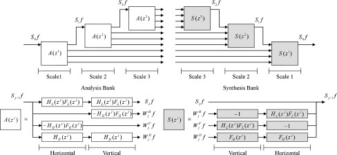
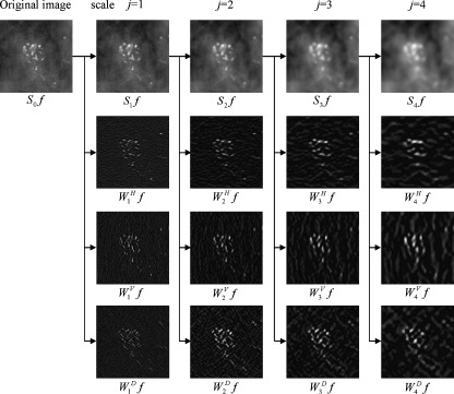
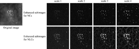
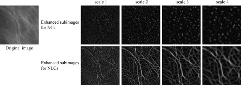
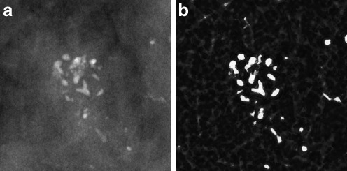
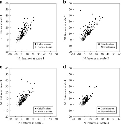
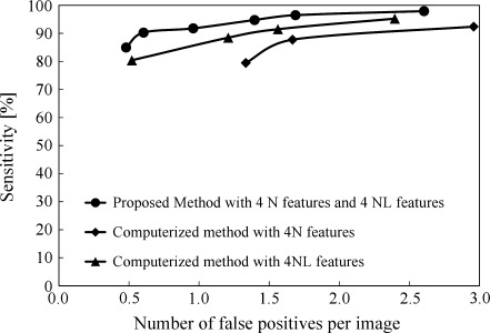
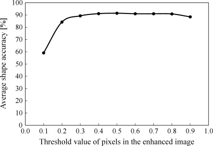
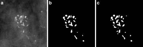
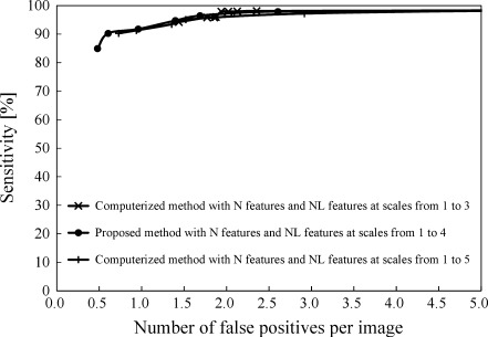
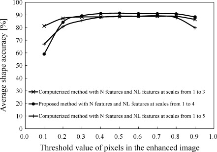
In a computer-aided diagnosis (CADx) scheme for evaluating the likelihood of malignancy of clustered microcalcifications on mammograms, it is necessary to segment individual calcifications correctly. The purpose of this study was to develop a computerized segmentation method for individual calcifications with various sizes while maintaining their shapes in the CADx schemes. Our database consisted of 96 magnification mammograms with 96 clustered microcalcifications. In our proposed method, a mammogram image was decomposed into horizontal subimages, vertical subimages, and diagonal subimages for a second difference at scales 1 to 4 by using a filter bank. The enhanced subimages for nodular components (NCs) and the enhanced subimages for both nodular and linear components (NLCs) were obtained from analysis of a Hessian matrix composed of the pixel values in those subimages for the second difference at each scale. At each pixel, eight objective features were given by pixel values in the subimages for NCs at scales 1 to 4 and the subimages for NLCs at scales 1 to 4. An artificial neural network with the eight objective features was employed to enhance calcifications on magnification mammograms. Calcifications were finally segmented by applying a gray-level thresholding technique to the enhanced image for calcifications. With the proposed method, a sensitivity of calcifications within clustered microcalcifications and the number of false positives per image were 96.5% (603/625) and 1.69, respectively. The average shape accuracy for segmented calcifications was also 91.4%. The proposed method with high sensitivity of calcifications while maintaining their shapes would be useful in the CADx schemes.
Figures

Similar articles
-
Computer-aided diagnosis scheme for histological classification of clustered microcalcifications on magnification mammograms.Med Phys. 2004 Apr;31(4):789-99. doi: 10.1118/1.1655711. Med Phys. 2004. PMID: 15124996
-
Computer-aided diagnosis scheme using a filter bank for detection of microcalcification clusters in mammograms.IEEE Trans Biomed Eng. 2006 Feb;53(2):273-83. doi: 10.1109/TBME.2005.862536. IEEE Trans Biomed Eng. 2006. PMID: 16485756
-
Segmentation of suspicious clustered microcalcifications in mammograms.Med Phys. 2000 Jan;27(1):13-22. doi: 10.1118/1.598852. Med Phys. 2000. PMID: 10659733
-
CADx of mammographic masses and clustered microcalcifications: a review.Med Phys. 2009 Jun;36(6):2052-68. doi: 10.1118/1.3121511. Med Phys. 2009. PMID: 19610294 Review.
-
[Computer aided diagnosis of calcifications in mammograms].Sheng Wu Yi Xue Gong Cheng Xue Za Zhi. 2011 Feb;28(1):170-4. Sheng Wu Yi Xue Gong Cheng Xue Za Zhi. 2011. PMID: 21485207 Review. Chinese.
Cited by
-
Methods on Skull Stripping of MRI Head Scan Images-a Review.J Digit Imaging. 2016 Jun;29(3):365-79. doi: 10.1007/s10278-015-9847-8. J Digit Imaging. 2016. PMID: 26628083 Free PMC article. Review.
-
Breast Cancer Segmentation Methods: Current Status and Future Potentials.Biomed Res Int. 2021 Jul 20;2021:9962109. doi: 10.1155/2021/9962109. eCollection 2021. Biomed Res Int. 2021. PMID: 34337066 Free PMC article. Review.
-
A Micro CT Study in Patients with Breast Microcalcifications Using a Mathematical Algorithm to Assess 3D Structure.PLoS One. 2017 Jan 20;12(1):e0169349. doi: 10.1371/journal.pone.0169349. eCollection 2017. PLoS One. 2017. PMID: 28107436 Free PMC article.
References
-
- Sickles EA. Mammographic features of early breast cancer. AJR AM J Roentgenol. 1984;143:461–464. - PubMed
-
- Sickles EA, Mammographic features of 300 consecutive nonpalpable breast cancers, AJR AM J Roentgenol, 661–663 (1986). - PubMed
-
- Adler D.D., Helvie M.A., Mammographic biopsy recommendations, Current Opinion in Radiology, 123–129 (1992). - PubMed
-
- Kopans DB. The positive predictive value of mammography. AJR AM J Roentgenol. 1992;158:521–526. - PubMed
MeSH terms
LinkOut - more resources
Full Text Sources
Medical
