. 2011 Nov;138(21):4597-608.
doi: 10.1242/dev.067736.

Zebrafish mnx1 controls cell fate choice in the developing endocrine pancreas

Affiliations

Zebrafish mnx1 controls cell fate choice in the developing endocrine pancreas

Gokhan Dalgin et al. Development. 2011 Nov.

Abstract

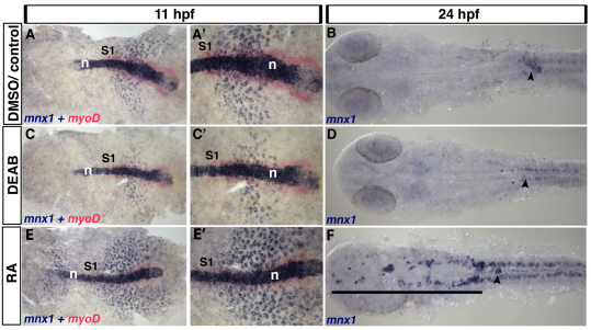
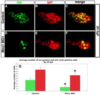
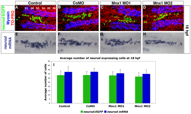
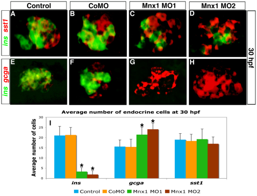
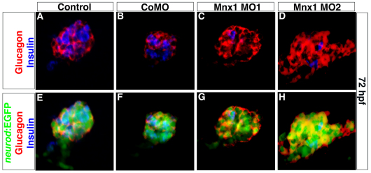
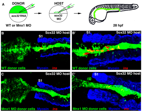
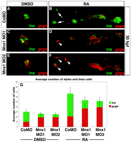
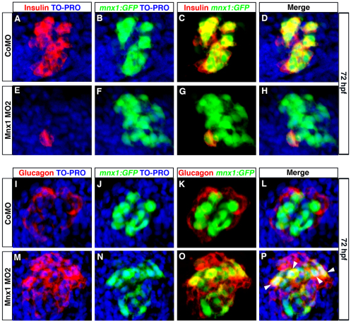
The vertebrate endocrine pancreas has the crucial function of maintaining blood sugar homeostasis. This role is dependent upon the development and maintenance of pancreatic islets comprising appropriate ratios of hormone-producing cells. In all vertebrate models studied, an initial precursor population of Pdx1-expressing endoderm cells gives rise to separate endocrine and exocrine cell lineages. Within the endocrine progenitor pool a variety of transcription factors influence cell fate decisions, such that hormone-producing differentiated cell types ultimately arise, including the insulin-producing beta cells and the antagonistically acting glucagon-producing alpha cells. In previous work, we established that the development of all pancreatic lineages requires retinoic acid (RA) signaling. We have used the zebrafish to uncover genes that function downstream of RA signaling, and here we identify mnx1 (hb9) as an RA-regulated endoderm transcription factor-encoding gene. By combining manipulation of gene function, cell transplantation approaches and transgenic reporter analysis we establish that Mnx1 functions downstream of RA within the endoderm to control cell fate decisions in the endocrine pancreas progenitor lineage. We confirm that Mnx1-deficient zebrafish lack beta cells, and, importantly, we make the novel observation that they concomitantly gain alpha cells. In Mnx1-deficient embryos, precursor cells that are normally destined to differentiate as beta cells instead take on an alpha cell fate. Our findings suggest that Mnx1 functions to promote beta and suppress alpha cell fates.


Figures

Fig. 1.
Fig. 1.
Endoderm expression of mnx1 is regulated by retinoic acid. (A-F) Control zebrafish embryos were treated with DMSO carrier (A,A′) or untreated (B). Retinoic acid (RA) signaling was blocked by DEAB treatment (C-D) or elevated by RA treatment (E-F). (A,C,E) In situ hybridization for mnx1 at 11 hpf shows punctate expression of mnx1 transcripts (blue) in the endoderm and high levels of expression in notochord. In situ hybridization for the mesodermal marker myod1 (myoD, red) marks the adaxial mesoderm at this stage up to the first somite (S1). (A′,C′,E′) Higher magnification views of A,C,E. At 24 hpf, mnx1 transcripts are localized to presumptive endocrine pancreas (B). DEAB treatment reduces the endodermal expression of mnx1 at 11 hpf (C,C′) and abolishes it at 24 hpf (arrowhead), whereas RA treatment increases the number of cells expressing mnx1 at 11 hpf (E,E′) and expands the mnx1-expressing pancreatic domain (arrowhead) towards the anterior (black line). Note that neither the notochord (A,C,E) nor the spinal cord motoneurons (B,D,F) show altered mnx1 expression in response to modulation of RA signaling. n, notochord.
Fig. 2.
Fig. 2.
Mnx1-deficient embryos have endocrine pancreas defects. (A-F) Confocal images of the dorsal pancreatic bud at 24 hpf. ins (green) and isl1 (red) transcripts were detected by double-fluorescent in situ hybridization (FISH) in control (A-C) or Mnx1 MO1-injected (D-F) zebrafish embryos. Anterior to left. (G) Mean (± s.d.) number of cells positive for insulin (ins) or isl1 (red) in control and mnx1 morphants. *, P<0.001 (t-test, two-tailed distribution, Bonferroni correction); control, n=35; Mnx1 MO1, n=50.
Fig. 3.
Fig. 3.
Endocrine progenitors are unaffected by Mnx1 knockdown. (A-D) Confocal images of Tg(neurod:EGFP) zebrafish embryos at 18 hpf. Whole-mount immunolabeling for GFP (green) and for myosin to label somites (blue); nuclear staining with TO-PRO-3 (red). Somites are numbered from anterior to posterior (S1-S6). (E-H) In situ hybridization for neurod at 18 hpf. (A,E) Uninjected controls; (B,F) control morphants; (C,G) Mnx1 MO1-injected embryos; (D,H) Mnx1 MO2-injected embryos. (I) Mean (± s.d.) neurod-expressing cells from two independent experiments and from a minimum of 17 embryos per group.
Fig. 4.
Fig. 4.
Mnx1-deficient embryos lack beta cells but gain alpha cells. (A-H) Confocal images of the dorsal pancreatic bud at 30 hpf showing double FISH for (A-D) ins (green) and somatostatin (sst1, red) or (E-H) ins (green) and glucagon (gcga, red) in zebrafish embryos that were uninjected (A,E) or injected with control MO (B,F), Mnx1 MO1 (C,G) or Mnx1 MO2 (D,H). (I) Mean (± s.d.) number of cells expressing the three markers. *, P<0.001 (t-test, two-tailed distribution, Bonferroni correction). Results are from three independent experiments with a minimum of ten embryos per group. CoMO, control MO.
Fig. 5.
Fig. 5.
The altered balance of alpha and beta cells in Mnx1-deficient embryos is maintained after ventral bud development. Confocal images of whole-mount 72 hpf Tg(neurod:EGFP) zebrafish embryos that were (A,E) uninjected, or injected with (B,F) control MO, (C,G) Mnx1 MO1 or (D,H) Mnx1 MO2, immunolabeled for glucagon (red), insulin (blue) and GFP (green) as indicated. Data are representative of three independent experiments.
Fig. 6.
Fig. 6.
Mnx1 functions directly in the endoderm to promote beta cell fate. (A) The cell transplantation approach. Wild-type zebrafish embryos were injected with fluorescein dextran (green) together with either sox32 mRNA or sox32 mRNA and Mnx1 MO. Control or morphant donor cells were transplanted into sox32 morphant hosts then raised until 28 hpf. (B-C′) Confocal images of representative 28 hpf control (B,B′) and Mnx1 MO (C,C′) transplants, at low (B,C) and higher (B′,C′) magnification, showing whole-mount immunostaining for myosin (blue) and in situ hybridization for insulin (red). S1, first somite. Anterior to left. Control, n=12; Mnx1 MO, n=13.
Fig. 7.
Fig. 7.
Mnx1 knockdown alters the alpha:beta cell ratio in a similar fashion in control and RA-treated embryos. (A-F) Confocal images of DMSO carrier-treated (A,C,E) and RA-treated (B,D,F) 30 hpf zebrafish embryos that were injected with control MO (A,B), Mnx1 MO1 (C,D) or Mnx1 MO2 (E,F), showing double FISH for ins (green) and gcga (red). Arrows indicate beta cells and arrowheads indicate alpha cells. (G) Mean (± s.d.) number of alpha and beta cells from four independent experiments with a minimum of 45 embryos per group. Anterior to left.
Fig. 8.
Fig. 8.
Beta cell progenitors take on an alpha cell fate in Mnx1-deficient embryos. (A-P) Confocal images of 72 hpf Tg(mnx1:GFP) zebrafish embryos injected with control MO (A-D,I-L) or Mnx1 MO2 (E-H,M-P). (A-H) Whole-mount immunolabeling for GFP (green) and insulin (red), with nuclear staining with TO-PRO-3 (blue). (I-P) Whole-mount immunostaining for GFP (green) and glucagon (red), with nuclear staining with TO-PRO-3 (blue). Arrowheads indicate colocalization of glucagon and GFP. Data are representative of three independent experiments with a minimum of 20 embryos per group.

References

    1. Anderson K. R., Torres C. A., Solomon K., Becker T. C., Newgard C. B., Wright C. V., Hagman J., Sussel L. (2009). Cooperative transcriptional regulation of the essential pancreatic islet gene NeuroD1 (beta2) by Nkx2.2 and neurogenin 3. J. Biol. Chem. 284, 31236-31248 - PMC - PubMed
    1. Argenton F., Zecchin E., Bortolussi M. (1999). Early appearance of pancreatic hormone-expressing cells in the zebrafish embryo. Mech. Dev. 87, 217-221 - PubMed
    1. Biemar F., Argenton F., Schmidtke R., Epperlein S., Peers B., Driever W. (2001). Pancreas development in zebrafish: early dispersed appearance of endocrine hormone expressing cells and their convergence to form the definitive islet. Dev. Biol. 230, 189-203 - PubMed
    1. Binot A.-C., Manfroid I., Flasse L., Winandy M., Motte P., Martial J. A., Peers B., Voz M. L. (2010). Nkx6.1 and nkx6.2 regulate alpha- and beta-cell formation in zebrafish by acting on pancreatic endocrine progenitor cells. Dev. Biol. 340, 397-407 - PubMed
    1. Chao C. S., Loomis Z. L., Lee J. E., Sussel L. (2007). Genetic identification of a novel NeuroD1 function in the early differentiation of islet alpha, PP and epsilon cells. Dev. Biol. 312, 523-532 - PMC - PubMed

Publication types

MeSH terms

LinkOut - more resources