Comparative Study
. 2012 Feb;35(2):399-408.
doi: 10.1002/jmri.22815. Epub 2011 Oct 11.

Fast Dixon whole-body MRI for detecting distant cancer metastasis: a preliminary clinical study

Affiliations
Comparative Study

Fast Dixon whole-body MRI for detecting distant cancer metastasis: a preliminary clinical study

Colleen M Costelloe et al. J Magn Reson Imaging. 2012 Feb.

Abstract

Purpose: To evaluate the feasibility of fast Dixon whole-body (WB) magnetic resonance imaging (MRI) for detecting bone and liver metastasis in clinical patients and to compare its performance with skeletal scintigraphy (SS) for detecting bone metastases using reference imaging with >1 year follow-up as the gold standard.

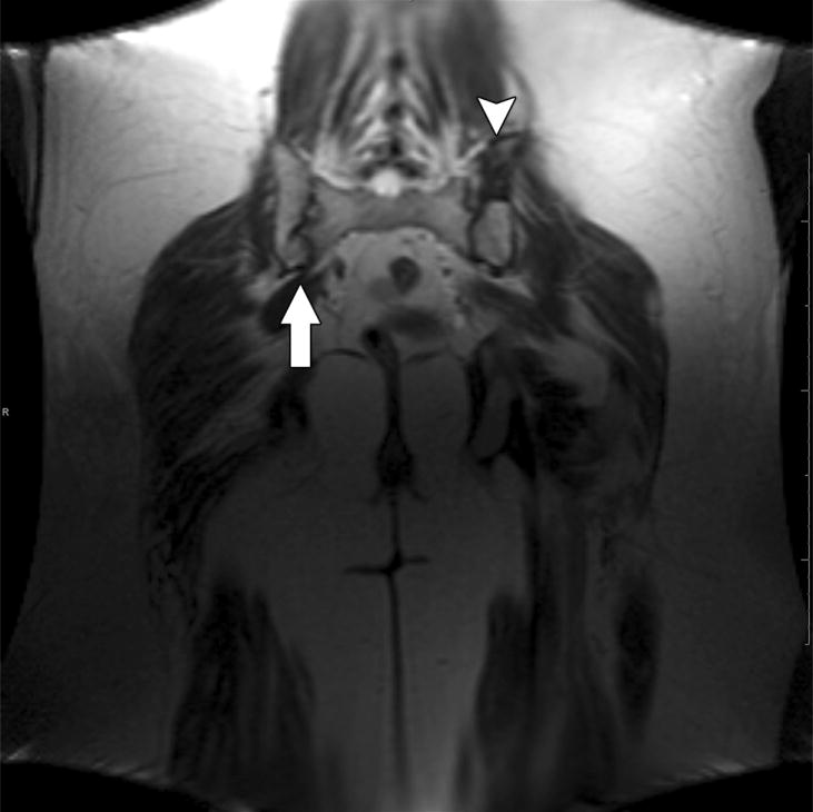
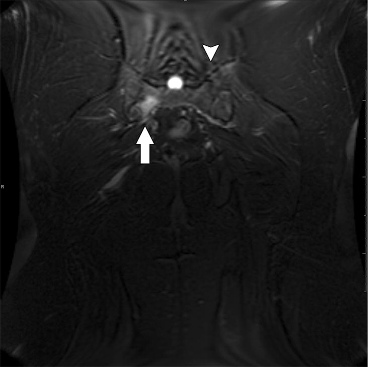
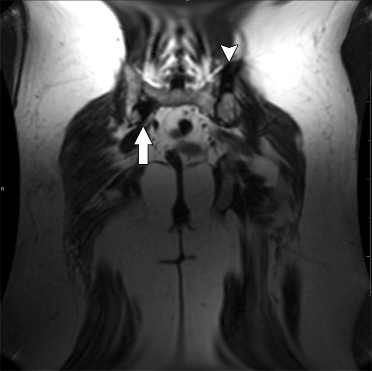
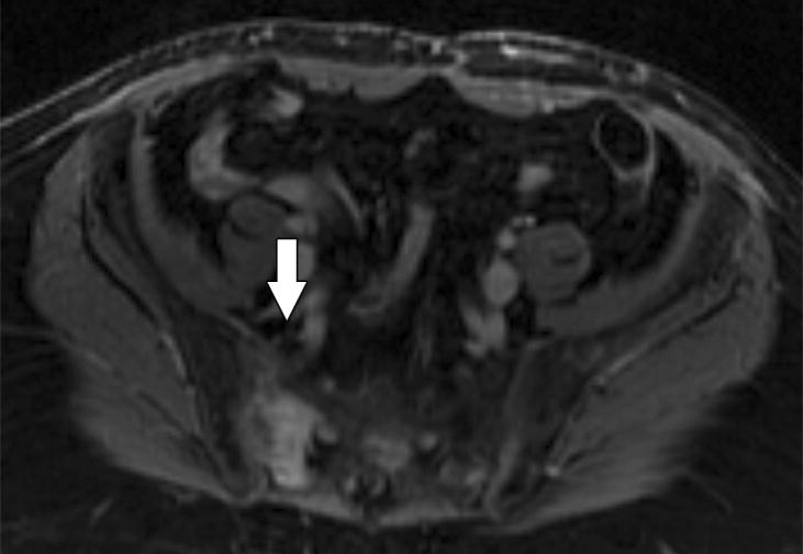
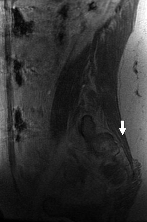
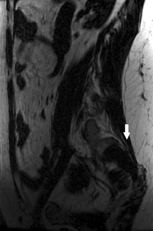
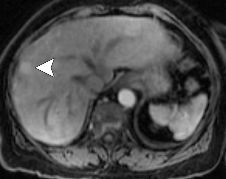
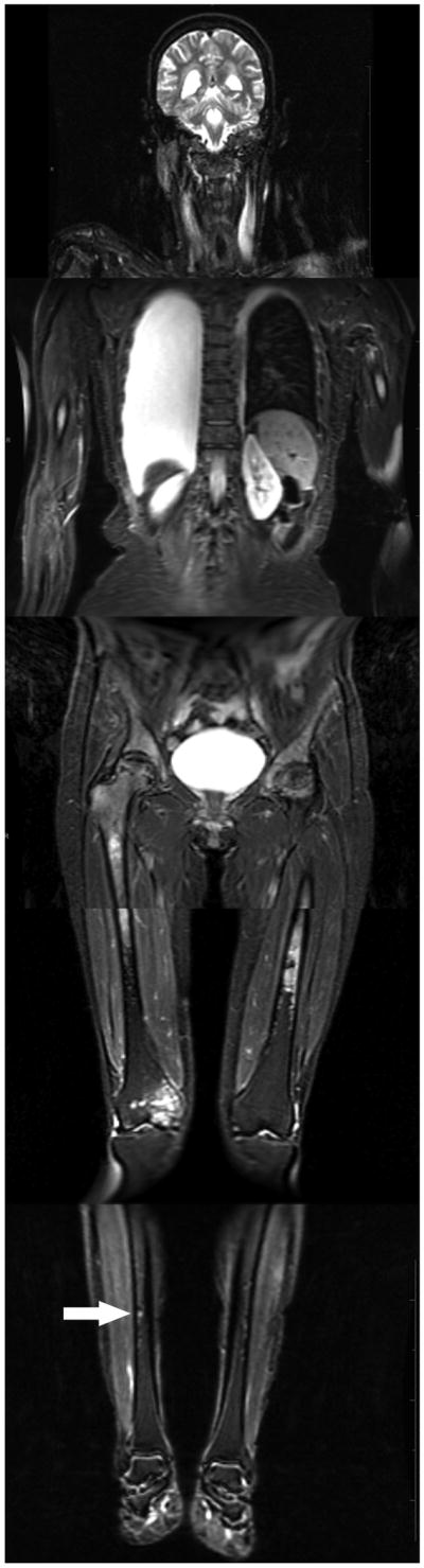
Materials and methods: Twenty-nine patients with bone metastases prospectively underwent WB MRI and SS. WB MRI included coronal T2, axial T1 with and without intravenous gadolinium (including triphasic liver sequences), and axial diffusion-weighted imaging, plus spinal sagittal postcontrast T1-weighted images. The skeleton was divided into 16 segments. Reviewers blinded to other images identified up to five lesions per segment and rated them using a five-point confidence scale for metastatic disease. Sensitivities and specificities were compared using the McNemar test.

Results: The sensitivity of WB MRI and SS in detecting bone metastases was 70.8% and 59.6% (P = 0.003), respectively; specificity was 89.1% and 98.7% (P < 0.0001). WB MRI detected all livers with metastases (n = 8). One focal nodular hyperplasia was classified as a metastasis on WB MRI.

Conclusion: Fast Dixon WB MRI is feasible in clinical patients, highly specific, and more sensitive than SS in detecting bone metastases, and can detect metastases of the liver.


Conflict of interest statement

Conflicts of Interest An author, Jingfei Ma, Ph.D, is the inventor of a United States of America patent on the fast Dixon sequences used in this study.

Figures

Figure 1
Figure 1
WB MRI can detect more bone metastasis than skeletal scintigraphy. (a) Axial FS T1 weighted image of the pelvis in a 58 year old female with metastatic breast cancer to bone. The diffuse high signal intensity indicates innumerable heterogeneously enhancing metastases. (b) Skeletal scintigraphy detected only one metastasis to the bony pelvis (arrow).
Figure 1
Figure 1
WB MRI can detect more bone metastasis than skeletal scintigraphy. (a) Axial FS T1 weighted image of the pelvis in a 58 year old female with metastatic breast cancer to bone. The diffuse high signal intensity indicates innumerable heterogeneously enhancing metastases. (b) Skeletal scintigraphy detected only one metastasis to the bony pelvis (arrow).
Figure 2
Figure 2
Diagram of the distribution of bone metastases of breast cancer by reference imaging. The numbers of metastases detected on reference images in each anatomic segment are followed in parentheses by the respective percentages of all metastases. The majority of the anatomic segments with more than five lesions had diffuse or innumerable metastases. These segments were considered to have six lesions, resulting in an underestimate of the total number of bone metastases. C spine, cervical spine; R, right; L, left; T spine, thoracic spine; L spine, lumbar spine.
Figure 3
Figure 3
Diagram of bone metastases of breast cancer per anatomic segment detected using WB MRI and bone scan. The numbers of bone metastases detected using WB MRI (blue) and skeletal scintigraphy (red) are followed in parentheses by the respective percentages of metastases shown on reference images per anatomic segment. Anatomical segments are shaded according to the modality that detected the larger proportion of lesions. The forearms and lower legs were excluded due to paucity of lesions. C spine, cervical spine; R, right; L, left; T spine, thoracic spine, L spine, lumbar spine.
Figure 4
Figure 4
Single session multisequence, multiplanar imaging of bone a metastasis. The fast Dixon protocol generated a large number of images that could seem overwhelming. This figure follows a single bone metastasis from a 43 year old breast cance patient through the majority of the images on which it was visible and shows how the large number of sequences and imaging planes complement each other to enhance diagnostic efficacy. (a-c) Dixon imaging produces 3 sequences per acquisition such as (a) coronal T2, (b) water only (fat-saturated) and (c) fat-only images of the pelvis. The lesion in the right sacral ala (arrow) is not conspicuous on (a) because of similar signal intensity to the surrounding fat. It is quite conspicuous on (b) due to high water content and can also be seen on (c) because it contains no fat. In contradistinction to the lesion in the right sacral ala, the lesion in the left iliac bone (arrowhead) is quite conspicuous on (a) because it has low T2 signal. It is not conspicuous on (b) due to low water content and is well seen on (c) due to lack of fat content. The differences in imaging characteristics between these 2 lesions emphasize the clinical usefulness of multisequence imaging for the detection of bone metastases. (d-i) The following illustrations will focus on the lesion in the right sacral ala which is at a different anatomic level than the lesion in the left iliac bone. Imaging in the axial plane was T1-weighted. (d) The lesion in the right sacral ala is low in signal intensity, (e) is higher than the surrounding fat on the water-only/fat-saturated image (f) and is low in signal intensity on the fat-only image. (g) Following contrast, the lesion enhances but is most conspicuous and easiest to detect on the (h) water-only image. (i) The two fat-only images are identical to visual assessment. (j-l) The spinal sagittal images were also post-contrast and were performed laterally enough to include the lesion in the right sacral ala. Signal characteristics were similar to the post-contrast axial images. (k) If the lesion was overlooked in the axial plane, the water-only, post-contrast image would likely aid in detection. (m) Lastly, the lesion demonstrated more restricted diffusion than surrounding structures, facilitating detection on the DWI sequence. Several lesions were identified only on the diffusion-weighted images (not shown). This illustration has been an example of the wide variety of sequences and imaging planes provided by the fast Dixon technique as per a lesion in the pelvis. Our protocol also included head/neck, chest/abdomen, thigh, calf/feet stations as well as the remainder of the spine in approximately one hour. The large number of sequences and imaging planes increases the likelihood that lesions with differing signal characteristics and locations will be detected.
Figure 4
Figure 4
Single session multisequence, multiplanar imaging of bone a metastasis. The fast Dixon protocol generated a large number of images that could seem overwhelming. This figure follows a single bone metastasis from a 43 year old breast cance patient through the majority of the images on which it was visible and shows how the large number of sequences and imaging planes complement each other to enhance diagnostic efficacy. (a-c) Dixon imaging produces 3 sequences per acquisition such as (a) coronal T2, (b) water only (fat-saturated) and (c) fat-only images of the pelvis. The lesion in the right sacral ala (arrow) is not conspicuous on (a) because of similar signal intensity to the surrounding fat. It is quite conspicuous on (b) due to high water content and can also be seen on (c) because it contains no fat. In contradistinction to the lesion in the right sacral ala, the lesion in the left iliac bone (arrowhead) is quite conspicuous on (a) because it has low T2 signal. It is not conspicuous on (b) due to low water content and is well seen on (c) due to lack of fat content. The differences in imaging characteristics between these 2 lesions emphasize the clinical usefulness of multisequence imaging for the detection of bone metastases. (d-i) The following illustrations will focus on the lesion in the right sacral ala which is at a different anatomic level than the lesion in the left iliac bone. Imaging in the axial plane was T1-weighted. (d) The lesion in the right sacral ala is low in signal intensity, (e) is higher than the surrounding fat on the water-only/fat-saturated image (f) and is low in signal intensity on the fat-only image. (g) Following contrast, the lesion enhances but is most conspicuous and easiest to detect on the (h) water-only image. (i) The two fat-only images are identical to visual assessment. (j-l) The spinal sagittal images were also post-contrast and were performed laterally enough to include the lesion in the right sacral ala. Signal characteristics were similar to the post-contrast axial images. (k) If the lesion was overlooked in the axial plane, the water-only, post-contrast image would likely aid in detection. (m) Lastly, the lesion demonstrated more restricted diffusion than surrounding structures, facilitating detection on the DWI sequence. Several lesions were identified only on the diffusion-weighted images (not shown). This illustration has been an example of the wide variety of sequences and imaging planes provided by the fast Dixon technique as per a lesion in the pelvis. Our protocol also included head/neck, chest/abdomen, thigh, calf/feet stations as well as the remainder of the spine in approximately one hour. The large number of sequences and imaging planes increases the likelihood that lesions with differing signal characteristics and locations will be detected.
Figure 5
Figure 5
Detection of liver metastasis on triphasic abdominal imaging. (a) Solid enhancement is seen in a liver lesion on the arterial phase (30 s following contrast administration, arrowhead). (b) Rapid contrast wash-out is evident on the 60 s and confirmed on the (c) 120 s images. The fast Dixon-based whole body protocol enabled the detection of metastases in multiple organ systems in a single imaging session.
Figure 5
Figure 5
Detection of liver metastasis on triphasic abdominal imaging. (a) Solid enhancement is seen in a liver lesion on the arterial phase (30 s following contrast administration, arrowhead). (b) Rapid contrast wash-out is evident on the 60 s and confirmed on the (c) 120 s images. The fast Dixon-based whole body protocol enabled the detection of metastases in multiple organ systems in a single imaging session.
Figure 6
Figure 6
Limitations of reference imaging for verifying bony metastases. (a) Composite coronal FS T2-weighted WB MRI scan demonstrating diffuse, high-signal-intensity bone metastases in the spine, humeri, pelvis, mid-proximal femora, and right distal femur (compared with the normal low signal of suppressed normal fat in the distal left femur and left tibia). A small, round focus of high signal intensity in the right tibial diaphysis (arrow) was rated with high confidence for metastatic disease. (b) Reference bone scan obtained 1 year after the WB MRI scan shown in (a) demonstrated no abnormality in the right tibia. Prior bone scans (not shown) also showed no abnormality in this area. No other reference imaging modality was available for the right tibial anatomic segment. Although the resolution of the right tibial lesion on the WB MRI scan was lower than the bone scan (7-12 mm), the WB MRI finding was considered a false-positive result. Situations such as this may have biased the specificity of WB MRI in our study. A large right and small left pleural effusion are also shown, demonstrating multi-organ system imaging with WB MRI.
Figure 6
Figure 6
Limitations of reference imaging for verifying bony metastases. (a) Composite coronal FS T2-weighted WB MRI scan demonstrating diffuse, high-signal-intensity bone metastases in the spine, humeri, pelvis, mid-proximal femora, and right distal femur (compared with the normal low signal of suppressed normal fat in the distal left femur and left tibia). A small, round focus of high signal intensity in the right tibial diaphysis (arrow) was rated with high confidence for metastatic disease. (b) Reference bone scan obtained 1 year after the WB MRI scan shown in (a) demonstrated no abnormality in the right tibia. Prior bone scans (not shown) also showed no abnormality in this area. No other reference imaging modality was available for the right tibial anatomic segment. Although the resolution of the right tibial lesion on the WB MRI scan was lower than the bone scan (7-12 mm), the WB MRI finding was considered a false-positive result. Situations such as this may have biased the specificity of WB MRI in our study. A large right and small left pleural effusion are also shown, demonstrating multi-organ system imaging with WB MRI.

References

    1. Abrams HL, Spiro R, Goldstein N. Metastases in carcinoma; analysis of 1000 autopsied cases. Cancer. 1950;3:74–85. - PubMed
    1. Costelloe CM, Rohren EM, Madewell JE, et al. Imaging bone metastases in breast cancer: techniques and recommendations for diagnosis. Lancet Oncol. 2009;10:606–614. - PubMed
    1. Horvath LJ, Burtness BA, McCarthy S, Johnson KM. Total-body echo-planar MR imaging in the staging of breast cancer: comparison with conventional methods--early experience. Radiology. 1999;211:119–128. - PubMed
    1. Engelhard K, Hollenbach HP, Wohlfart K, von Imhoff E, Fellner FA. Comparison of whole-body MRI with automatic moving table technique and bone scintigraphy for screening for bone metastases in patients with breast cancer. Eur Radiol. 2004;14:99–105. - PubMed
    1. Lauenstein TC, Goehde SC, Herborn CU, et al. Whole-body MR imaging: evaluation of patients for metastases. Radiology. 2004;233:139–148. - PubMed

Publication types