. 2012 May;72(5):743-52.
doi: 10.1002/dneu.20987.

The pubertal-related decline in cellular proliferation and neurogenesis in the dentate gyrus of male rats is independent of the pubertal rise in gonadal hormones

Affiliations

The pubertal-related decline in cellular proliferation and neurogenesis in the dentate gyrus of male rats is independent of the pubertal rise in gonadal hormones

Amy Ho et al. Dev Neurobiol. 2012 May.

Abstract

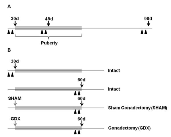
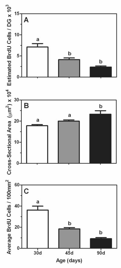
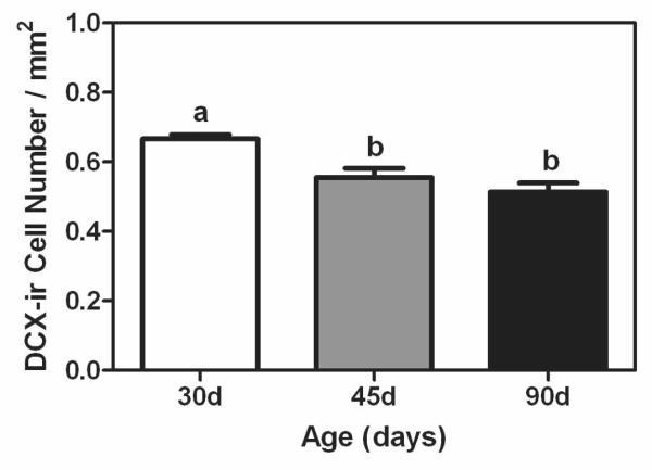
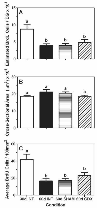
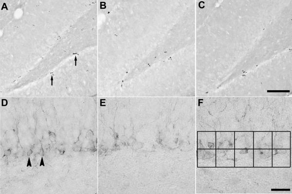
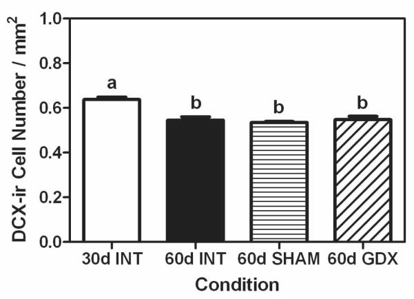
Pubertal development is marked by significant decreases in cellular proliferation and neurogenesis in the dentate gyrus of the hippocampal formation. Although it is unclear what mediates these developmental changes in the dentate gyrus, gonadal hormones have been implicated in modulating many neurobiological processes during puberty and various parameters of neurogenesis in adulthood. Thus, it is possible that the gradual and sustained increase in gonadal hormones experienced during puberty plays a role in these changes in neurogenesis. In this experiments, we first quantified cellular proliferation and neurogenesis using 5-bromo-2'-deoxyuridine (BrdU) and doublecortin (DCX) immunohistochemistry, respectively, in the dentate gyrus of prepubertal (30 d), midpubertal (45 d), and adult (90 d) male rats. We found the decline in BrdU and DCX cell numbers throughout these ages was coincident with increases in their plasma testosterone levels. We next tested whether exposure to the pubertal rise in gonadal hormones was necessary for this decrease in hippocampal neurogenesis to occur. Thus, we examined cellular proliferation and neurogenesis in intact 30 day (prepubertal) and 60-day-old (late-pubertal) rats, as well as 60-day-old rats that had previously been gonadectomized or sham-gonadectomized at 30 days of age. Although we again found the expected decline in BrdU and DCX cell numbers between 30 and 60 days of age in the intact groups, there were no differences among the 60-day-old animals, regardless of gonadal status. These data indicate that the pubertal-related decline in hippocampal cellular proliferation and neurogenesis is independent of the pubertal change in gonadal hormones.


Figures

Figure 1
Figure 1
Schematic timelines for the experimental designs in experiments 1 (A) and 2 (B). Black arrows indicate the day tissues were collected, gray arrows the day of surgery, and black arrowheads the BrdU injections (200 mg/kg). The gray bar on each timeline demarcates the approximate time span of pubertal development. Abbreviations: d, days of age; GDX, gonadectomy; SHAM, sham-gonadectomy.
Figure 2
Figure 2
Mean (±SEM) estimated number of BrdU-positive cells (A), average cross-sectional area (μm2; B), and average number of BrdU-positive cells per 100mm2 (C) in the dentate gyrus of prepubertal (30 days of age, white bars), mid-pubertal (45 days of age, gray bars), and adult (90 days of age, black bars) male rats. Bars that share the same letter are not significantly different from each other.
Figure 3
Figure 3
Mean (±SEM) number of DCX-positive cells per mm2 in dentate gyrus of prepubertal (30 days of age, white bar), mid-pubertal (45 days of age, gray bar), and adult (90 days of age, black bar) male rats. Bars that share the same letter are not significantly different from each other.
Figure 4
Figure 4
Mean (±SEM) estimated number of BrdU-positive cells (A), average cross-sectional area (μm2; B), and average number of BrdU-positive cell number per 100mm2 (C) in dentate gyrus of intact prepubertal (30 days of age INT, white bars), intact late-pubertal (60 days of age INT, black bars), sham-gonadectomized late-pubertal (SHAM, 60 days of age, bars with horizontal hash lines), and gonadectomized (GDX, 60 days of age, bars with diagonal hash lines) male rats. Bars that share the same letter are not significantly different from each other.
Figure 5
Figure 5
Representative photomicrographs of BrdU- and DCX-positive cells in the dentate gyrus. Photomicrographs on the top row represent BrdU-positive cells in (A) intact 30 day old males, (B) intact 60 day old males, and (C) 60 day old males that had been gonadectomized at 30 days of age. Photomicrographs on the bottom row represent DCX-positive cells in (D) intact 30 day old males, (E) intact 60 day old males, and (F) 60 day old males that had been gonadectomized at 30 days of age. Arrows in panel A are indicating examples of clusters of BrdU-positive cells, while arrowheads in panel D are indicating examples of DCX-positive cells. The black squares in panel F represent the approximate area (6,250μm2) and placement of the ocular grid used to quantify DCX-positive cell numbers. Scale bars = 100μm and 25μm for the top and bottom row of photomicrographs, respectively.
Figure 6
Figure 6
Mean (±SEM) number of DCX-positive cells per mm2 in dentate gyrus of prepubertal (30 days of age INT, white bars), intact late-pubertal (60 days of age INT, black bars), sham-gonadectomized late-pubertal (SHAM, 60 days of age, bars with horizontal hash lines), and gonadectomized (GDX, 60 days of age, bars with diagonal hash lines) male rats. Bars that share the same letter are not significantly different from each other.

Similar articles

Cited by

References

    1. Ahmed EI, Zehr JL, Schulz KM, Lorenz BH, DonCarlos LL, Sisk CL. Pubertal hormones modulate the addition of new cells to sexually dimorphic brain regions. Nat Neurosci. 2008;11:995–997. - PMC - PubMed
    1. Andersen SL, Thompson RF, Krenzel E, Teicher MH. Pubertal changes in gonadal hormones do not underlie adolescent dopamine receptor overproduction. Psychoneuroendocrinology. 2002;27:683–691. - PubMed
    1. Barker JM, Galea LAM. Repeated estradiol administration alters different aspects of neurogenesis and cell death in the hippocampus of female, but not male, rats. Neuroscience. 2008;152:888–902. - PubMed
    1. Buwalda B, van der Borght K, Koolhaas JM, McEwen BS. Testosterone decrease does not play a major role in the suppression of hippocampal cell proliferation following social defeat stress in rats. Physiol Behav. 2010;101:719–725. - PubMed
    1. Casey BJ, Duhoux S, Cohen MM. Adolescence: what do transmission, transition, and translation have to do with it? Neuron. 2010;67:749–760. - PMC - PubMed

Publication types