. 2011 Dec;152(12):5029-40.
doi: 10.1210/en.2011-1124. Epub 2011 Oct 11.

Lipopolysaccharide initiates inflammation in bovine granulosa cells via the TLR4 pathway and perturbs oocyte meiotic progression in vitro

Affiliations

Lipopolysaccharide initiates inflammation in bovine granulosa cells via the TLR4 pathway and perturbs oocyte meiotic progression in vitro

John J Bromfield et al. Endocrinology. 2011 Dec.

Abstract

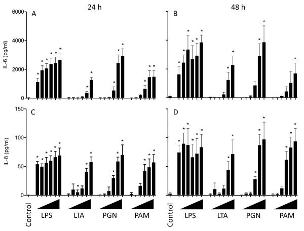
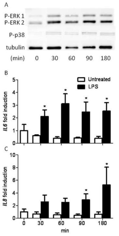
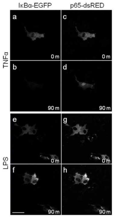
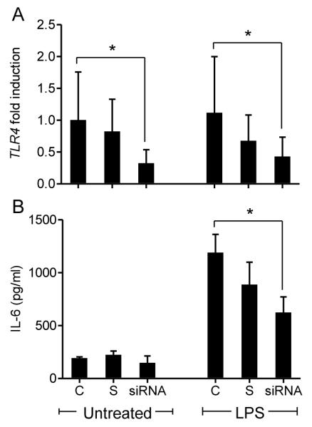
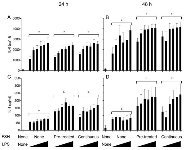
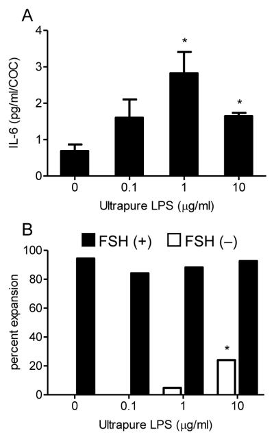
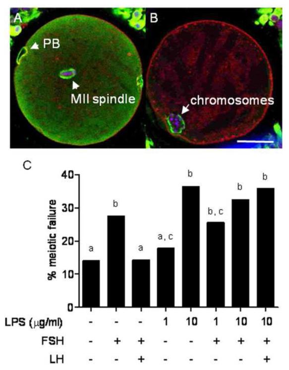
Infections of the reproductive tract or mammary gland with Gram-negative bacteria perturb ovarian function, follicular growth, and fecundity in cattle. We hypothesized that lipopolysaccharide (LPS) from Gram-negative bacteria stimulates an inflammatory response by ovarian granulosa cells that is mediated by Toll-like receptor (TLR) 4. The present study tested the capacity of bovine ovarian granulosa cells to initiate an inflammatory response to pathogen-associated molecular patterns and determined subsequent effects on the in vitro maturation of oocytes. Granulosa cells elicited an inflammatory response to pathogen-associated molecular patterns (LPS, lipoteichoic acid, peptidoglycan, or Pam3CSK4) with accumulation of the cytokine IL-6, and the chemokine IL-8, in a time- and dose-dependent manner. Granulosa cells responded acutely to LPS with rapid phosphorylation of TLR signaling components, p38 and ERK, and increased expression of IL6 and IL8 mRNA, although nuclear translocation of p65 was not evident. Targeting TLR4 with small interfering RNA attenuated granulosa cell accumulation of IL-6 in response to LPS. Endocrine function of granulosa cells is regulated by FSH, but here, FSH also enhanced responsiveness to LPS, increasing IL-6 and IL-8 accumulation. Furthermore, LPS stimulated IL-6 secretion and expansion by cumulus-oocyte complexes and increased rates of meiotic arrest and germinal vesicle breakdown failure. In conclusion, bovine granulosa cells initiate an innate immune response to LPS via the TLR4 pathway, leading to inflammation and to perturbation of meiotic competence.


Figures

Figure 1
Figure 1. Granulosa cell cultures are free of professional cells
Cultured granulosa cells were assessed for contamination with professional immune cells. (A) PCR amplification of MHCII and AMH in cultured granulosa and blood cells. (B) Confocal micrographs of cultured granulosa (a) and blood (b) cells showing MHCII immunoreactivity (green), DNA (blue) and DIC (a’, b’). (C) flow cytometry histograms of freshly isolated blood and granulosa cells showing MHC reactivity in red and unstained cells in grey. (D) Confocal micrographs representing a cross section of a 4-8 mm follicle (a), a <4 mm follicle (b) and spleen (c) showing MHCII immunoreactivity (green), DNA (blue) and DIC (a’, b’, c’). Arrow heads indicate MHCII positive cells, the asterix represents the antral cavity and the dashed line represents the basement membrane extrapolated from the DIC image. Scale bar represents 20 μm.
Figure 2
Figure 2. Granulosa cells produced IL-6 and IL-8 in response to LPS, LTA, PGN and PAM in a dose-dependent manner
Accumulation of IL-6 (A, B) and IL-8 (C, D) in the supernatants of granulosa cells was measured by ELISA following 24 h (A, C) or 48 h (B, D) in culture with the PAMPs ultrapure lipopolysaccharide (LPS), lipoteichoic acid (LTA), peptidoglycan (PGN) or Pam3CSK4 (PAM). Granulosa cells were treated with 10-fold increasing doses of each PAMP from 100 pg/ml up to 10 μg/ml of each PAMP (ranging left to right and represented by triangles). Data are presented as mean + SEM from 4 independent experiments. * P < 0.05 compared to untreated controls; analysis by ANOVA followed by Dunnett’s pair-wise post-hoc tests.
Figure 3
Figure 3. LPS induced phosphorylation of ERK and p38, and IL6 and IL8 gene expression in granulosa cells in a time-dependent manner
(A) Granulosa cells were treated with 1 μg/ml of ultrapure LPS and cultured for 30, 60, 90 or 180 min before Western blot analysis. Bands are shown corresponding to di-phosphorylated ERK 1/2, phosphorylated p38 (Thr180/Tyr182) and tubulin as a loading control. Image is representative of 3 independent experiments. Granulosa cells were cultured in control medium (□) or medium containing 1 μg/ml of ultrapure LPS (■) for 30, 60, 90 or 180 min and the IL6 (B) and IL8 (C) gene expression was measured using real time RT-PCR. Data are represented as mean + SEM fold induction compared with control cells at time 0 from 4 independent experiments. * P < 0.05 compared to untreated control within time points; analysis by non-parametric Mann-Whitney U test.
Figure 4
Figure 4. Nuclear translocation of p65 or IκBα degradation in granulosa cells was induced by TNFα but not LPS
Granulosa cells were cultured in glass bottom petri dishes and transfected with IκBα-EGFP (a-b, e-f) and p65-dsRed (c-d, e-h). Live cell confocal microscopy under culture conditions was performed to track degradation of IκBα-EGFP and nuclear translocation of p65-dsRed. Cells were treated with 20 ng/ml TNFα (a-d) as a positive control, or 1 μg/ml ultrapure LPS (e-h). Scale bar represents 50μm.
Figure 5
Figure 5. siRNA targeting of TLR4 in granulosa cells reduced TLR4 gene expression and the accumulation of IL-6 in response to LPS
Granulosa cells were cultured for 24 h with lipofectamine (control, C), scramble-siRNA (S) or a specific TLR4-targeted siRNA (siRNA) prior to treatment with 1 μg/ml of ultrapure LPS for an additional 24 h. (A) Real time RT-PCR was used to evaluate the knock down of TLR4 mRNA with expression presented as fold-change relative to untreated controls. (B) IL-6 accumulation was measured in cell-free supernatants by ELSIA following 24 h treatment with 1 μg/ml of ultrapure LPS. Data are presented as mean + SEM from 4 independent experiments. * P < 0.05 compared to controls within treatment group. Real time RT-PCR data were analyzed using a non-parametric Mann-Whitney U test, ELISA data were analysed using ANOVA followed by a Bonferonni post-hoc test.
Figure 6
Figure 6. Exogenous FSH increases accumulation of IL-6 and IL-8 in response to LPS by granulosa cells
Accumulation of IL-6 (A, B) and IL-8 (C, D) measured by ELISA following 24 h (A, C) or 48 h (B, D) in culture after challenge of granulosa cells with ultrapure LPS. Granulosa cells were either pre-incubated with 2.5 μg/ml FSH for 6 h prior to LPS exposure or treated with 2.5 μg/ml FSH for the duration of the LPS exposure. Granulosa cells were challenged with 10-fold increasing doses of ultrapure LPS between 100 pg/ml and 10 μg/ml (ranging left to right and represented by triangles). Data are presented as mean + SEM from 4 independent experiments. Different superscripts represent statistically significant differences between FSH treatment groups (P < 0.05); analysis by ANOVA followed by Bonferonni post-hoc test.
Figure 7
Figure 7. LPS induces IL-6 accumulation and expansion in in vitro matured COCs
(A) IL-6 accumulation in cell-free supernatants was measured from groups of 10 – 20 COCs treated with 0, 0.1, 1 or 10 μg/ml LPS in the presence of 2.5 μg/ml FSH. IL-6 concentrations are presented as mean pg/ml/COC + SEM from a total of 216 across 4 independent experiments. * P < 0.05 compared to untreated controls; analysis by ANOVA followed by Dunnett’s post-hoc test. (B) COC expansion rates were recorded from groups of 10 – 20 COCs,, following treatment with 0, 0.1, 1 or 10 μg/ml LPS in the absence (□) or presence (■) of 2.5 μg/ml FSH. COC expansion data are presented as percentages from a total of 422 COCs across 8 independent experiment. * P < 0.05 compared to untreated COCs within FSH treatment group using a χ2 test.
Figure 8
Figure 8. LPS induces meiotic failure in in vitro matured COCs
Meiotic progression was assessed in 290 COCs by confocal microscopy. Oocytes at the M-phase of meiosis with a normal bipolar spindle and aligned chromosomes were deemed to have completed meiosis (A). Any oocyte not to reach the M-phase of meiosis II or those which had significantly perturbed meiotic structures such as aberrant spindles, chromosomal ejection, parthenogenic activation or germinal vesicle breakdown failure were deemed to have failed meiosis (B). Representative confocal micrographs of COCs show DNA (blue), tubulin (green) and F-actin (red). (C) Meiotic failure rates of COCs cultured in the defined IVM media containing LPS, FSH and or LH (as indicated) for 24 h are presented as percentages. Different superscripts represent statistical significance following a χ2 test; P < 0.05. PB, polar body; scale bar represents 50 μm.

References

    1. Sheldon IM, Noakes DE, Rycroft AN, Pfeiffer DU, Dobson H. Influence of uterine bacterial contamination after parturition on ovarian dominant follicle selection and follicle growth and function in cattle. Reproduction. 2002;123:837–845. - PubMed
    1. Herath S, Williams EJ, Lilly ST, Gilbert RO, Dobson H, Bryant CE, Sheldon IM. Ovarian follicular cells have innate immune capabilities that modulate their endocrine function. Reproduction. 2007;134:683–693. - PMC - PubMed
    1. Williams EJ, Sibley K, Miller AN, Lane EA, Fishwick J, Nash DM, Herath S, England GC, Dobson H, Sheldon IM. The effect of Escherichia coli lipopolysaccharide and tumour necrosis factor alpha on ovarian function. Am J Reprod Immunol. 2008;60:462–473. - PMC - PubMed
    1. Lavon Y, Leitner G, Klipper E, Moallem U, Meidan R, Wolfenson D. Subclinical, chronic intramammary infection lowers steroid concentrations and gene expression in bovine preovulatory follicles. Domest Anim Endocrinol. 2011;40:98–109. - PubMed
    1. Dohmen MJ, Joop K, Sturk A, Bols PE, Lohuis JA. Relationship between intrauterine bacterial contamination, endotoxin levels and the development of endometritis in postpartum cows with dystocia or retained placenta. Theriogenology. 2000;54:1019–1032. - PubMed

Publication types