. 2011 Oct 25;108(43):17815-20.
doi: 10.1073/pnas.1110655108. Epub 2011 Oct 11.

Glial-conditional deletion of aquaporin-4 (Aqp4) reduces blood-brain water uptake and confers barrier function on perivascular astrocyte endfeet

Affiliations

Glial-conditional deletion of aquaporin-4 (Aqp4) reduces blood-brain water uptake and confers barrier function on perivascular astrocyte endfeet

Nadia Nabil Haj-Yasein et al. Proc Natl Acad Sci U S A. .

Abstract

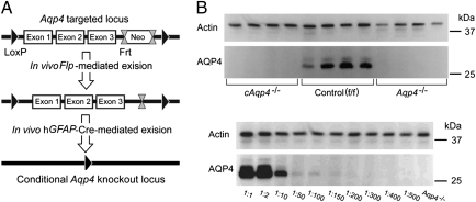
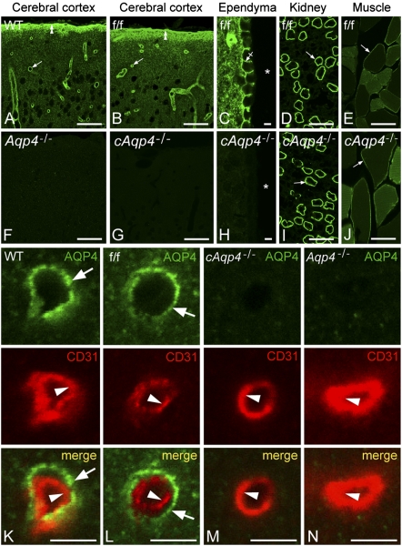
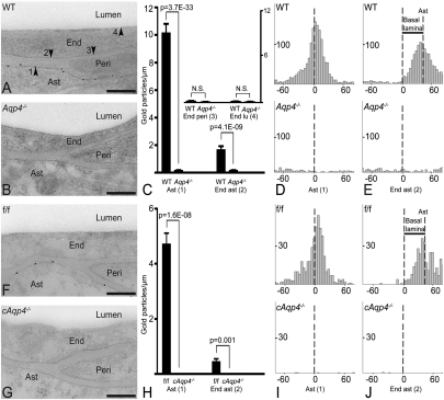
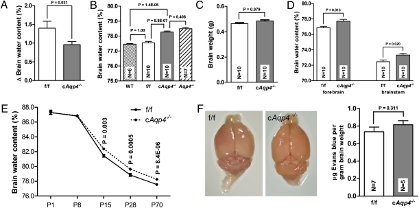
Tissue- and cell-specific deletion of the Aqp4 gene is required to differentiate between the numerous pools of aquaporin-4 (AQP4) water channels. A glial-conditional Aqp4 knockout mouse line was generated to resolve whether astroglial AQP4 controls water exchange across the blood-brain interface. The conditional knockout was driven by the glial fibrillary acidic protein promoter. Brains from conditional Aqp4 knockouts were devoid of AQP4 as assessed by Western blots, ruling out the presence of a significant endothelial pool of AQP4. In agreement, immunofluorescence analysis of cryostate sections and quantitative immunogold analysis of ultrathin sections revealed no AQP4 signals in capillary endothelia. Compared with litter controls, glial-conditional Aqp4 knockout mice showed a 31% reduction in brain water uptake after systemic hypoosmotic stress and a delayed postnatal resorption of brain water. Deletion of astroglial Aqp4 did not affect the barrier function to macromolecules. Our data suggest that the blood-brain barrier (BBB) is more complex than anticipated. Notably, under certain conditions, the astrocyte covering of brain microvessels is rate limiting to water movement.


Conflict of interest statement

The authors declare no conflict of interest.

Figures

Fig. 1.
Fig. 1.
Strategy for generation and validation of glial-conditional Aqp4 knockout mice. (A) FRT-neomycin-FRT-LoxP validated cassette was inserted downstream of exon (Ex) 3, and a LoxP site was inserted upstream of exon 1. The Neo cassette was deleted by breeding chimeras with Flp recombinase-expressing mice. Heterozygous Aqp4 floxed mice were bred with mice expressing Cre recombinase under the human glial fibrillary acidic protein (hGFAP) promoter, and after intercrossing offspring, conditional Aqp4−/− (cAqp4−/−) mice were obtained. (B, Upper) Immunoblots of brain homogenates obtained from four cAqp4−/− mice (lanes 1–4), four litter f/f control mice (lanes 5–8), and four constitutive Aqp4−/−mice (lanes 9–12). The blots were probed with anti-actin (for loading control) and anti-AQP4 antibodies, as indicated to the Left. The AQP4 blot showed two confluent bands of ∼32 kDa and 34 kDa in homogenates from f/f control mice. These bands were absent in homogenates from both glial-conditional and constitutive Aqp4−/−mice, which were indistinguishable. (Lower) Immunoblots of WT brain homogenates diluted in homogenates from constitutive Aqp4−/−mice. The blots were probed with antibodies against actin and AQP4, respectively, and show that AQP4 immunosignals easily could be detected when 1% or more of the homogenate originated from WT mice. Actin staining verifies similar loading. The 25-kDa and 37-kDa markers are indicated on the Right.
Fig. 2.
Fig. 2.
Immunofluorescence micrographs showing distribution of AQP4. The AQP4 labeling (green) pattern in cerebrum of WT (A) and f/f mice (B and C) was indistinguishable. Note distinct immunosignal around vessels (arrows), underneath the pia (double arrowheads), and along the basolateral membrane of ependymocytes (crossed arrow). Asterisks, third ventricle. In cerebrum of constitutive (F) and cAqp4−/− (G and H) mice, AQP4 immunoreactivity was absent. Conditional deletion of Aqp4, however, did not affect the labeling pattern in muscle and kidney (compare I and J with D and F; arrows indicate kidney collecting duct and sarcolemma, respectively). Double labeling of obliquely cut capillaries with the endothelial marker CD31 (red) revealed that the AQP4 labeling (green) associated with microvessels in WT and f/f mice was peripheral to the endothelium (arrowhead), corresponding to perivascular astrocytic endfeet (arrows; K and L). No detectable signal was seen over endothelial cells in either genotype (K and N). (Scale bars: A, B, D, E, F, G, I, and J, 50 μm; C, H, and KN, 5 μm.)
Fig. 3.
Fig. 3.
Subcellular distribution of AQP4 immunogold reactivity. (A) Electron micrograph from WT mouse showing AQP4 immunogold particles along the astrocyte (Ast) endfoot membrane. End, endothelial cell; Peri, pericyte. Arrowheads indicate astrocyte endfoot membrane (1) and the endothelial membranes facing astrocyte (2), pericyte (3), and lumen (4). (B) AQP4 labeling was absent in constitutive Aqp4 knockout mice (Aqp4−/− mice). (C) Quantitative analysis of the density of gold particles over the membrane domains indicated in A. The same symbols (14) are used for easy reference. Gold particles within 23.5 nm from a given membrane were allocated to that membrane. The AQP4 signal over the Ast endfoot membrane was significantly higher in WT than in Aqp4−/− mice. The two genotypes did not differ in AQP4 densities over endothelial membranes facing pericytes (End peri) and lumen (End lu), indicating absence of AQP4 in these membrane domains. The AQP4 signal over endothelial membranes next to astrocyte endfeet (End ast) was higher in WT than in Aqp4−/− mice. (D and E, Upper) Analysis in WT animals of the distribution of gold particles perpendicular to the astrocyte endfoot membrane (Ast, D) or to the endothelial membrane facing astrocytes (End ast, E) revealed peak immunogold signal coinciding with former membrane. When analysis was performed with abluminal endothelial membrane as reference (E) the peak was skewed corresponding to the thickness of the basal lamina (indicated with bar; Results). (D and E, Lower) No peaks were seen in Aqp4−/− mice. (F) AQP4 immunogold labeling pattern in controls (f/f) was similar to that of WT mice (A). (G) AQP4 immunoreactivity was absent in glial-conditional Aqp4 knockout mice (cAqp4−/− mice). (H) The linear densities of AQP4 signaling gold particles over astrocytic and apposed endothelial membranes showed a pronounced reduction in cAqp4−/− vs. litter controls (f/f). (I and J) Same design as in D and E, respectively, but f/f controls in lieu of WT and cAqp4−/− mice in lieu of Aqp4−/− mice. Perpendicular distribution of gold particles showed absence of peaks after glial-conditional deletion of Aqp4, confirming that the immunosignals seen in f/f mice reside in glia. (Scale bars: A, B, F, and G, 200 nm.)
Fig. 4.
Fig. 4.
Effect of glial-conditional Aqp4 deletion on brain water content and blood–brain barrier permeability. (A) Increase in brain water content 40 min after i.p. water injection (150 mL/kg) was significantly lower in cAqp4−/− mice than in f/f litter controls. Error bars indicate SE. (B) Basal brain water content of cAqp4−/− mice was significantly higher than that of WT and f/f controls, but did not differ from that of constitutive Aqp4−/− mice. (C) Brain mass did not differ significantly between genotypes (P > 0.05). (D) Baseline water content of forebrain hemispheres and brainstem was higher in cAQP4−/− mice than in controls. (E) Baseline brain water content at various stages of postnatal development. At postnatal days 1 (P1) and 8 (P8) the genotypes did not differ. At P15, P28, and P70, the brain water content of cAqp4−/− mice was significantly higher than that of f/f mice. (F) Blood–brain barrier macromolecule permeability. Evans blue staining was not observed in f/f or cAqp4−/− mouse brains subsequent to i.v. injection. No significant difference was observed in mean Evans blue content between the two genotypes.

References

    1. Bechmann I, Galea I, Perry VH. What is the blood-brain barrier (not)? Trends Immunol. 2007;28:5–11. - PubMed
    1. Abbott NJ, Rönnbäck L, Hansson E. Astrocyte-endothelial interactions at the blood-brain barrier. Nat Rev Neurosci. 2006;7:41–53. - PubMed
    1. Reese TS, Karnovsky MJ. Fine structural localization of a blood-brain barrier to exogenous peroxidase. J Cell Biol. 1967;34:207–217. - PMC - PubMed
    1. Manley GT, et al. Aquaporin-4 deletion in mice reduces brain edema after acute water intoxication and ischemic stroke. Nat Med. 2000;6:159–163. - PubMed
    1. Nielsen S, et al. Specialized membrane domains for water transport in glial cells: High-resolution immunogold cytochemistry of aquaporin-4 in rat brain. J Neurosci. 1997;17:171–180. - PMC - PubMed

Publication types

LinkOut - more resources