. 2011 Dec 9;286(49):42180-42187.
doi: 10.1074/jbc.M111.298711. Epub 2011 Oct 11.

S1 pocket of a bacterially derived subtilisin-like protease underpins effective tissue destruction

Affiliations

S1 pocket of a bacterially derived subtilisin-like protease underpins effective tissue destruction

Wilson Wong et al. J Biol Chem. .

Abstract

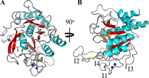
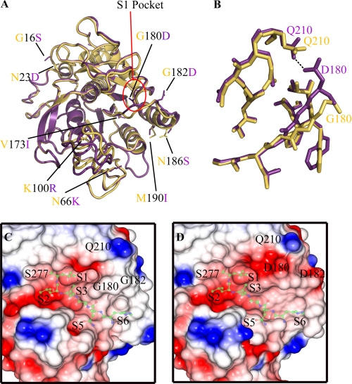
The ovine footrot pathogen, Dichelobacter nodosus, secretes three subtilisin-like proteases that play an important role in the pathogenesis of footrot through their ability to mediate tissue destruction. Virulent and benign strains of D. nodosus secrete the basic proteases BprV and BprB, respectively, with the catalytic domain of these enzymes having 96% sequence identity. At present, it is not known how sequence variation between these two putative virulence factors influences their respective biological activity. We have determined the high resolution crystal structures of BprV and BprB. These data reveal that that the S1 pocket of BprV is more hydrophobic but smaller than that of BprB. We show that BprV is more effective than BprB in degrading extracellular matrix components of the host tissue. Mutation of two residues around the S1 pocket of BprB to the equivalent residues in BprV dramatically enhanced its proteolytic activity against elastin substrates. Application of a novel approach for profiling substrate specificity, the Rapid Endopeptidase Profiling Library (REPLi) method, revealed that both enzymes prefer cleaving after hydrophobic residues (and in particular P1 leucine) but that BprV has more restricted primary substrate specificity than BprB. Furthermore, for P1 Leu-containing substrates we found that BprV is a significantly more efficient enzyme than BprB. Collectively, these data illuminate how subtle changes in D. nodosus proteases may significantly influence tissue destruction as part of the ovine footrot pathogenesis process.


Figures

FIGURE 1.
FIGURE 1.
1.8 Å crystal structure of BprV. A, view of the BprV structure showing the conserved subtilisin-like fold. Disulfide bonds are shown as yellow sticks, and the three calcium ions are shown as blue spheres. B, view of BprV structure with the I1 to I4 loops labeled. The figure was prepared using PyMOL (31).
FIGURE 2.
FIGURE 2.
Structural variations between the BprV and BprB proteases. A, overlay between the BprV and BprB structures showing the location of the nine amino acid substitutions. BprV-specific residues are shown as yellow sticks, and BprB-specific residues are shown as purple sticks. The S1 pockets of the proteases are highlighted by a red circle. B, structural overlay of the residues that form the S1 pockets of BprV and BprB. The S1 pockets of BprV and BprB are shown as yellow and purple sticks, respectively. The S1 pockets of both proteases are formed by residues 176–180, 204–208, and 215–218. C and D, electrostatic potential surfaces of the S1 pockets of BprV (C) and BprB (D) showing the Gly180 and Gly182 residues of BprV and Asp180 and Asp182 residues of BprB. The catalytic residue Ser277 is labeled, as is the Gln210 residue with which Asp180 of BprB makes a hydrogen bond (shown). Neutral regions are colored white, acidic regions are colored red, and basic regions are colored blue. Semitransparent peptide substrates (ball and stick) have been modeled onto the substrate binding clefts of BprV and BprB and are included to help visualize the subsites of the two proteases. A and B were prepared using PyMOL (31), and C and D were prepared using CCP4MG (32, 33).
FIGURE 3.
FIGURE 3.
Protease activity of BprV (V) and BprB (B) on components of the extracellular matrix. A, 50 nm purified protease incubated with 1 μm fibronectin at 25 °C. Samples were taken at the indicated time points, and the reaction was stopped by the addition of SDS-PAGE loading buffer and subsequent boiling. Degraded fibronectin products were analyzed using SDS-PAGE. B, quantitative measurement of fibronectin cleavage by densitometry analysis. C, quantitative measurement of elastase activity of the basic proteases. Each purified protease (5 μm) was incubated with 5 mg of Congo Red elastin at 37 °C for 5 h. Elastase activity was measured by reading the absorbance of the recovered supernatant at 490 nm for detection of the proteolytically released Congo Red dye. Mean ± S.E. (error bars) from triplicate experiments are shown, and statistical significance was calculated using one-way ANOVA (p < 0.05 between BprV and BprB, p < 0.05 for all BprB mutants compared with BprB). D, activities of BprV and BprB on ovine hoof material tested by incubating 100 μg/ml of the proteases with a 14% (w/v) insoluble hoof fragment at 37 °C. Samples of supernatant were taken at the indicated time points, and the reaction was stopped by the addition of SDS-PAGE loading buffer, followed by boiling for 5 min. Degraded hoof products were analyzed using SDS-PAGE. The 12-kDa band (asterisk) corresponding to the major degraded product was excised and subjected to in-gel trypsin digestion, followed by LC-MS analysis, to determine the identity of degraded product. E, quantitative measurement of keratin cleavage by densitometry analysis.
FIGURE 4.
FIGURE 4.
Identification of REPLi peptides cleaved by the BprV and BprB proteases. A, rate of cleavage of eight REPLi peptide pools best cleaved by the BprV protease. The peptide pools (50 μm) were incubated with 10 nm BprV at 37 °C. Fluorescence intensities were measured at 30-s intervals for 30 min. The initial velocities of cleavage for the pools are shown. B, rate of cleavage of eight synthesized peptides based on the (I/L)-(A/V)-(F/Y) pool of the REPLi library by BprV and BprB. The proteases (70 nm) were incubated with each peptide substrate (50 μm) at 37 °C, and fluorescence intensities were measured at 30-s intervals for 30 min. The initial velocities of cleavage of each peptide are shown. Mean ± S.E. (error bars) from a triplicate experiment are shown. C, representative mass spectra identifying cleaved products of the peptides, LAY, LAF, LVY, and IAF after cleavage with BprV and BprB.

Similar articles

Cited by

References

    1. Roberts D. S., Egerton J. R. (1969) J. Comp. Pathol. 79, 217–227 - PubMed
    1. Green L. E., George T. R. (2008) Vet. J. 175, 173–180 - PubMed
    1. Stewart D. J. (1989) in Footrot and Foot Abscess of Ruminants (Egerton J. R., Yong W. K., Riffkin G. G. eds) pp. 5–45, CRC Press, Boca Raton, FL
    1. Wani S. A., Samanta I. (2006) Vet. J. 171, 421–428 - PubMed
    1. Billington S. J., Johnston J. L., Rood J. I. (1996) FEMS Microbiol. Lett. 145, 147–156 - PubMed

Publication types

LinkOut - more resources