. 2011 Oct;23(10):3641-53.
doi: 10.1105/tpc.111.090944. Epub 2011 Oct 11.

INDEHISCENT and SPATULA interact to specify carpel and valve margin tissue and thus promote seed dispersal in Arabidopsis

Affiliations

INDEHISCENT and SPATULA interact to specify carpel and valve margin tissue and thus promote seed dispersal in Arabidopsis

Thomas Girin et al. Plant Cell. 2011 Oct.

Abstract

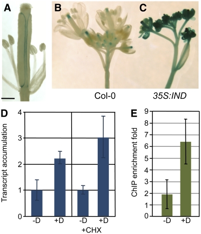
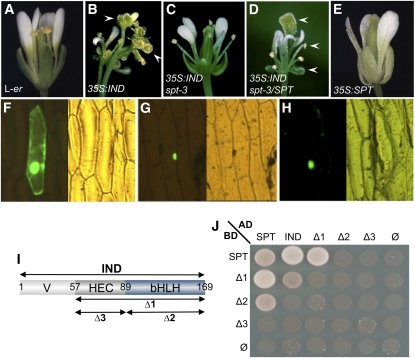
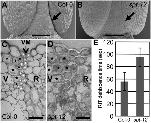
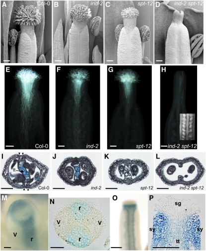
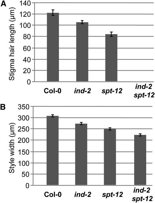
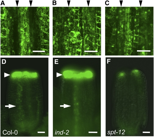
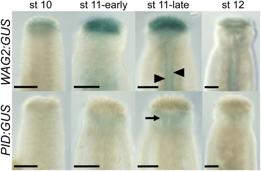
Structural organization of organs in multicellular organisms occurs through intricate patterning mechanisms that often involve complex interactions between transcription factors in regulatory networks. For example, INDEHISCENT (IND), a basic helix-loop-helix (bHLH) transcription factor, specifies formation of the narrow stripes of valve margin tissue, where Arabidopsis thaliana fruits open on maturity. Another bHLH transcription factor, SPATULA (SPT), is required for reproductive tissue development from carpel margins in the Arabidopsis gynoecium before fertilization. Previous studies have therefore assigned the function of SPT to early gynoecium stages and IND to later fruit stages of reproductive development. Here we report that these two transcription factors interact genetically and via protein-protein contact to mediate both gynoecium development and fruit opening. We show that IND directly and positively regulates the expression of SPT, and that spt mutants have partial defects in valve margin formation. Careful analysis of ind mutant gynoecia revealed slight defects in apical tissue formation, and combining mutations in IND and SPT dramatically enhanced both single-mutant phenotypes. Our data show that SPT and IND at least partially mediate their joint functions in gynoecium and fruit development by controlling auxin distribution and suggest that this occurs through cooperative binding to regulatory sequences in downstream target genes.


Figures

Figure 1.
Figure 1.
SPT Expression Is Directly Induced by IND. (A) Expression of a SPT:GUS reporter construct (−1262 bp) at the valve margin of a stage-16 Arabidopsis fruit (Smyth et al. 1990). (B) and (C) SPT:GUS (−6253 bp) expression in inflorescences of Col-0 (B) and 35S:IND (C). (D) Q-RT-PCR analysis of SPT relative transcript accumulation after control treatment (−D) or IND activation by Dex treatment (+D) in a 35S:IND:GR line, in presence or absence of Chxe (+CHX). Values are the average of three biological repeats ± sd. +Dex values are significantly different from their corresponding −Dex-treated control (Student’s t test P value < 0.05). (E) Fold-enrichment of the SPT promoter region in immunoprecipitated chromatin using an anti-GR antibody on the 35S:IND:GR line after control treatment (−D) or Dex treatment (+D). Values are the average of four biological repeats ± sd. The values are significantly different (Student’s t test P value = 0.01). Bar in (A) = 0.5 mm.
Figure 2.
Figure 2.
Ectopic IND Function Is SPT-Dependent, and IND and SPT Proteins Interact. (A) to (E) Flowers of L-er (A), 35S:IND (B), 35S:IND spt-3 (C), 35S:IND spt-3/SPT (D), and 35S:SPT (E). Plants in (C) and (D) derive from the same transformation event. Arrowheads in (B) and (D) indicate ectopic stigma. (F) to (H) GFP localization in epidermal onion cells transiently transformed with 35S:SPTΔNLS:GFP alone (F), 35S:SPT (G), or 35S:IND (H). (I) Schematic of IND truncated versions used in (J). Variable N-terminal domain (V), the HEC-conserved domain (HEC), and the bHLH domain (bHLH) are indicated. Amino acid positions are shown. Δ1 construct contains HEC and bHLH domain, Δ2 contains the bHLH domain and Δ3 contains the HEC domain. (J) Yeast two-hybrid experiment using fusions of SPT and IND (full-length and truncated version) with GAL4 activation and binding domains (AD and BD, respectively). Cells were spotted on selective medium lacking Leu, Trp, His, and adenine.
Figure 3.
Figure 3.
SPT Is Involved in Valve Margin Specification. (A) and (B) Scanning electron microscopy of the base of Col-0 and spt-12 fruits at stage 18. Arrows indicate the dehiscence zone. (C) and (D) Transmission electron micrographs of valve margin (VM) region in Col-0 and spt-12 siliques (stage 17b). Black and white stars indicate cells from the lignified and separation layers of the VM, respectively. Valves (V), VM, and replum (R) regions are indicated. (E) Dehiscence assessment (Arabidopsis Random Impact Test) of Col-0 and spt-12. Values correspond to the time of shaking required to open 50% of dried siliques. Values are the average of at least three biological repeats (20 mature siliques for each) ± sd. The values are significantly different (Student’s t test P value = 0.02). Bars in (A) and (B) = 100 μm; bars in (C) and (D) = 10 μm.
Figure 4.
Figure 4.
IND and SPT Promote Formation of Marginal Tissues. (A) to (D) Scanning electron microscopy images of gynoecia apical tissues at stage 13 in Col-0, ind-2, spt-12, and ind-2 spt-12. (E) to (H) Pollen-tube growth in Col-0 (E), ind-2 (F), spt-12 (G), and ind-2 spt-12 (H). Inset in (H) is a light microscope image of ovules in an ind-2 spt-12 gynoecium. (I) to (L) Cross sections of stage-13 ovaries in the wild type (Col-0) (I), ind-2 (J), spt-12 (K), and ind-2 spt-12 (L). Tissues are indicated in the wild-type section (I): v, valve; r, replum; tt, transmitting tract that has been stained with Alcian Blue. (M) and (N) IND:IND:GUS expression in whole mount (M) and cross section (N) of stage-9 gynoecia. Presumptuous valves (v) and repla (r) are indicated. (O) and (P) IND:IND:GUS expression in whole mount (O) and longitudinal section (P) of stage-12 gynoecia. Stigma (sg), style (sy), and transmitting tract (tt) are indicated in (P). Bars in (A) to (H), (I) to (L), (O), and (P) = 100 μm; bars in (M) and (N) = 25 μm.
Figure 5.
Figure 5.
IND and SPT Regulate Stigma Hair Length and Style Width. (A) Length of stigma hairs in Col-0, ind-2, spt-12, and ind-2 spt-12. (B) Width of style in Col-0, ind-2, spt-12, and ind-2 spt-12. Values in (A) and (B) are the average of >28 measurements ± se. All the values in individual panels (A) and (B) are significantly different to each other (Student’s t test P value < 0.01).
Figure 6.
Figure 6.
IND and SPT Regulate Auxin Distribution. (A) to (C) DR5:GFP expression in VM region of stage-17b fruits of Col-0 (A), ind-2 (B), and spt-12 (C). Black arrowheads above the images indicate the position of VM creases, which show a gap in fluorescence in the wild-type but not in the ind-2 and spt-12 fruits. (D) to (F) DR5:GFP expression in stage-9 gynoecia of Col-0 (D), ind-2 (E), and spt-12 (F). Arrowheads and arrows indicate respectively GFP signals at the top of the gynoecium and in the presumptive replum. Bars in (A) to (C) = 50 μm; bars in (D) to (F) = 25 μm.
Figure 7.
Figure 7.
Expression of WAG2 and PID in the Developing Gynoecium. Histochemical staining of gynoecia from WAG2:GUS (Top) and PID:GUS (Bottom) reporter lines throughout development. Developmental stages are indicated above. Arrowheads in WAG2:GUS (st 11-late) indicate expression in the medial region, and arrow in PID:GUS (st 11-late) points to weak expression in the style. Bars = 50 μm.
Figure 8.
Figure 8.
IND and SPT Cooperate to Regulate Downstream Targets and Bind Separate but Adjacent Elements in the Promoter of PINOID. (A) Scanning electron microscopy of style and stigmatic tissues in pid-9. (B) Quantification of yeast one-hybrid interactions between ALC, IND, and SPT proteins and two different elements of PID promoter (E-box variant and Double G-box). ALC was used as a negative control (nonspecific binding of a bHLH protein). Values are averages of five replicates ± sd. Asterisk (*) indicates values that are significantly different from the corresponding ALC control (Student’s t test P value < 0.0001). (C) and (D) Q-PCR analysis of PID (C) and WAG2 (D) relative to transcript accumulation after control treatment (−D) or IND activation (+D) in 35S:IND:GR (SPT) or 35S:IND:GR spt-12 (spt-12). Values are the average of at least four biological repeats ± se. Asterisk (*) indicates values that are significantly different from their corresponding −Dex-treated control (Student’s t test P value < 0.02). (E) Schematic of SPT and IND binding to the PID promoter. The double G-box elements are orange and the E-box is green. TATA indicates the position of the transcription initiation TATA box, and the PID coding region is shown in red. Bar in (A) = 100 μm.

Similar articles

Cited by

References

    1. Alvarez J., Smyth D.R. (1999). CRABS CLAW and SPATULA, two Arabidopsis genes that control carpel development in parallel with AGAMOUS. Development 126: 2377–2386 - PubMed
    1. Alvarez J., Smyth D.R. (2002). CRABS CLAW and SPATULA genes regulate growth and pattern formation during gynoecium development in Arabidopsis thaliana. Int. J. Plant Sci. 163: 17–41
    1. Alvarez J.P., Goldshmidt A., Efroni I., Bowman J.L., Eshed Y. (2009). The NGATHA distal organ development genes are essential for style specification in Arabidopsis. Plant Cell 21: 1373–1393 - PMC - PubMed
    1. Arnaud N., Girin T., Sorefan K., Fuentes S., Wood T.A., Lawrenson T., Sablowski R., Østergaard L. (2010). Gibberellins control fruit patterning in Arabidopsis thaliana. Genes Dev. 24: 2127–2132 - PMC - PubMed
    1. Balanzá V., Navarrete M., Trigueros M., Ferrándiz C. (2006). Patterning the female side of Arabidopsis: The importance of hormones. J. Exp. Bot. 57: 3457–3469 - PubMed

Publication types

MeSH terms

Substances

LinkOut - more resources