. 2011;6(10):e25641.
doi: 10.1371/journal.pone.0025641. Epub 2011 Oct 3.

RSPO1/β-catenin signaling pathway regulates oogonia differentiation and entry into meiosis in the mouse fetal ovary

Affiliations

RSPO1/β-catenin signaling pathway regulates oogonia differentiation and entry into meiosis in the mouse fetal ovary

Anne-Amandine Chassot et al. PLoS One. 2011.

Abstract

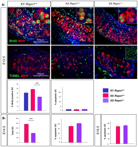
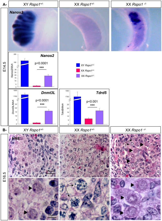
Differentiation of germ cells into male gonocytes or female oocytes is a central event in sexual reproduction. Proliferation and differentiation of fetal germ cells depend on the sex of the embryo. In male mouse embryos, germ cell proliferation is regulated by the RNA helicase Mouse Vasa homolog gene and factors synthesized by the somatic Sertoli cells promote gonocyte differentiation. In the female, ovarian differentiation requires activation of the WNT/β-catenin signaling pathway in the somatic cells by the secreted protein RSPO1. Using mouse models, we now show that Rspo1 also activates the WNT/β-catenin signaling pathway in germ cells. In XX Rspo1(-/-) gonads, germ cell proliferation, expression of the early meiotic marker Stra8, and entry into meiosis are all impaired. In these gonads, impaired entry into meiosis and germ cell sex reversal occur prior to detectable Sertoli cell differentiation, suggesting that β-catenin signaling acts within the germ cells to promote oogonial differentiation and entry into meiosis. Our results demonstrate that RSPO1/β-catenin signaling is involved in meiosis in fetal germ cells and contributes to the cellular decision of germ cells to differentiate into oocyte or sperm.


Conflict of interest statement

Competing Interests: The authors have declared that no competing interests exist.

Figures

Figure 1
Figure 1. Rspo1 promotes XX germ cells proliferation.
A- Reduction of germ cell proliferation in XX Rspo1 mutant gonads. Immunodetection of the proliferating germ cells with BrdU and MVH (upper panel), and the apoptotic germ cells with TUNEL and MVH (middle panel), in XY and XX Rspo+/−, and XX Rspo1−/− gonads at E12.5 gonads. DAPI (blue): nuclei. Inset middle panel: positive control for TUNEL. Histograms: Percentage of proliferating germ cells or apoptotic germ cells in XY, XX Rspo1+/− and Rspo1−/− gonads at E12.5. Bars represent mean+1 SEM, n = 24 sections of each genotype. B- Ablation of Rspo1 does not trigger germ cell apopotosis. Quantification of germ cell number and germ cell apoptosis in XX Rspo1+/− and Rspo1−/− gonads at E14.5 and E16.5. Bars represent mean+1 SEM, n = 24 sections of each genotype.
Figure 2
Figure 2. Rspo1 promotes XX germ cells meiosis.
Upper panel: Oct4 expression is maintained in XX Rspo1 mutant gonads. Oct4 whole-mount in situ hybridization at E14.5 in XY, XX control and XX Rspo1−/− fetal gonads. Middle panels: Downregulation of meiotic markers in XX Rspo1 mutant gonads. Immunodetection of SCP3 in E14.5 gonads and of γH2AX and MVH in E16.5 gonads. DAPI (blue): nuclei. Lower panel: Quantification of meiotic germ cells in XX Rspo1+/− and Rspo1−/− gonads at E14.5 (SCP3) and E16.5 (γH2AX). Bars represent mean+1 SEM, n = 15 sections of each genotype.
Figure 3
Figure 3. Rspo1 is involved in induction of Stra8 expression.
A- Quantitative RT-PCR analysis of Stra8, Cyp26b1, RARα and RARβ expression in E12.5 XY control, XX control, and Rspo1−/− gonads, using Mvh (for Stra8 qPCR) or Hprt (for other qPCR) as the normalization control. Bars represent mean+1 SEM, n = 8 individual embryos. B- In situ hybridizations at E14.5 in XY and XX control and XX Rspo1−/− gonads for Stra8 and Cyp26b1. C- In situ hybridizations on sections of the same XX Rspo1−/− gonad for Oct4 and Stra8 at E14.5.
Figure 4
Figure 4. Up-regulation of of gonocyte markers in XX Rspo1 mutant gonads.
A- In situ hybridizations at E14.5 in XY and XX control and XX Rspo1−/− gonads for Nanos2. Histograms: Quantitative RT-PCR analysis of Nanos2, Dnmt3L and Tdrd5 expression in E14.5 XY control, XX control and Rspo1−/− gonads, using Hprt as the normalization control. Bars represent mean+1 SEM, n = 8 individual embryos. B- Haematoxylin & Eosin staining of gonadic sections of XY, XX Rspo1+/− and XX Rspo1−/− gonads at E16.5. Meiotic oogonia are indicated by arrows GO/G1 quiescent gonocytes are indicated by arrowheads.
Figure 5
Figure 5. Germ cell sex reversal is not caused by Sertoli cell factors in XX Rspo1 mutant gonads.
A- Lack of Sertoli cell differentiation at E14.5 in XX Rspo1 mutant gonads. In situ hybridizations of Sox9 and Pgds in E14.5 XY and XX control and XX Rspo1−/− gonads. Right panels: Quantitative RT-PCR analysis of Sox9 and Pgds, using Hprt as the normalization control. Bars represent mean+1 SEM, n = 8 individual embryos. B- Fgf9 is not significantly expressed at E11.5 and E12.5 in XX Rspo1 mutant gonads. Upper panels: Quantitative RT-PCR analysis of Fgf9 at E11.5, E12.5 and E14.5 using Hprt as the normalization control. NS: Not significant. Bars represent mean+1 SEM, n = 8 individual embryos. Lower panels: In situ hybridizations of Fgf9 (E12.5 and E14.5) in XY and XX control and XX Rspo1−/− gonads.
Figure 6
Figure 6. Rspo1 activates β-catenin signaling pathway in XX germ cells.
A- Stabilization of β-catenin in XX germ cells is mediated by Rspo1. Active β-catenin (red) and MVH immunostaining (germ cells, green) in XY, XX Rspo1+/− and Rspo1−/− gonads at E14.5. DAPI (blue): nuclei. Arrowheads: nuclear β-catenin staining. B- Effectors of the WNT/β-catenin signaling pathway are expressed in XX germ cells. Quantitative RT-PCR analysis of Axin2, Lef1, LRP6 expression in E13.5 germ cells (XX and XY Oct4-positive cells) and somatic cells (XX and XY Oct4-negative cells), using respectively Hprt as the normalization control. Bars represent mean+1 SEM, n = 3 individual experiments. C- Expression of Axin2, a target of β-catenin in XX germ cells. X-Gal staining (AXIN2) (blue) and MVH immunostaining (germ cells, red) in XY Axin2+/LacZ, XX Axin2+/LacZ and XX Axin2+/LacZ; Rspo1−/− gonads at E12.5. Arrowheads: germ cells. G: gonad. X-Gal staining (AXIN2) (blue) and MVH immunostaining (germ cells, green) on isolated germ cells from XY Axin2+/LacZ and XX Axin2+/LacZ gonads at E12.5. Arrowheads: germ cells, arrows: somatic cells.
Figure 7
Figure 7. Activation of β-catenin promotes XY germ cell proliferation in fetal gonads.
A- Upper panel: Immunodetection of active β-catenin and MVH in XY and XX control and XY Catnbex3/+; TNAP:CreTr gonads at E14.5. Middle panel: Immunodetection of the proliferating germ cells with BrdU and MVH in XY and XX control, and XY Catnbex3/+; TNAP:CreTr gonads at E14.5. DAPI (blue): nuclei. Histograms: Percentage of BrdU-positive germ cells in XY and XX control, and XY Catnbex3/+; TNAP:CreTr gonads at E14.5. Bars represent mean+1 SEM, n = 24 sections of each genotype. Lower panel: Haematoxylin & Eosin staining of E15.5 gonadic sections from XY and XX wild type gonads and XY Catnbex3/+; TNAP:CreTr gonads. Arrowheads: XY quiescent gonocytes, arrows: meiotic germ cells, dashed arrows: mitotic germ cells, thin arrowhead: putative early meiotic germ cell. Histogram: Quantitative RT-PCR analysis of Stra8 at E15.5 using Mvh as the normalization control. Bars represent mean+1 SEM, n = 8 individual embryos. B- Upper panel: Immunostaining of FOXL2 (green) and E-Cadherin (germ cells, red) in XY and XX control, and XY Catnbex3/+; TNAP:CreTr gonads at E14.5. The white star indicates fluorescent blood cells within the coelomic vessel of the XY gonads. Lower panel: Immunodetection of the Sertoli cell marker SDMG1 in the same gonads. DAPI (blue): nuclei.
Figure 8
Figure 8. Somatic ablation of β-catenin does not affect germ cell fate.
A- Proliferation is not impaired in XX Catnb flox/flox; Sf1:CreTr gonads. Immunodetection of the proliferating germ cells with BrdU (green) and MVH (red) in XY, XX control and XX Catnb flox/flox; Sf1:CreTr gonads at E12.5. DAPI (blue): nuclei. Histograms: Percentage of BrdU-positive germ cells in XY, XX control and XX Catnb flox/flox; Sf1:CreTr gonads at E12.5. Bars represent mean+1 SEM, n = 24 sections of each genotype. B- Meiosis in not hampered in XXCatnb flox/flox; Sf1:CreTr gonads. Haematoxylin & Eosin staining of sections from E17.5 XY, XX control and XX Catnb flox/flox; Sf1:CreTr gonads. Arrowheads: gonocytes, arrows: meiotic germ cells, dotted arrows: apoptotic germ cells. Histograms: Percentage of apoptotic germ cells versus total germ cells after TUNEL staining in XY, XX control gonads and XX Catnb flox/flox; Sf1:CreTr gonads. Bars represent mean+1 SEM, n = 24 sections of each genotype. C: Immunodetection of SOX9 (red) in XY, XX control gonads and XX Catnb flox/flox; Sf1:CreTr gonads at E18.5. DAPI (blue) was used to detect nuclei.
Figure 9
Figure 9. Opposing signals regulate germ cell sexual differentiation.
Male and female fetal germ cells have acquired independent mechanisms to regulate their differentiation. In the fetal testis, Mvh is required to promote germ cell proliferation. FGF9 secreted by Sertoli cells promotes germ cell survival and may inhibit entry into meiosis. Then Nanos2 promotes differentiation into gonocytes and meiosis inhibition. RSPO1 has been shown to be a secreted activator of β-catenin. In fetal ovary, RSPO1/β-catenin signalling pathway promotes germ cell proliferation. Then, in addition to the retinoic acid (RA) signaling pathway, RSPO1 contributes in meiosis entry as a parallel pathway (indicated by the presence of two parallel arrows). RSPO1 promotes Stra8 expression and subsequently meiosis initiation. Although we showed that RSPO1 is a direct activator of β-catenin in XX germ cells suggesting that RSPO1 is also acting via β-catenin in this developmental process, the direct inactivation of β-catenin in XX germ cells remains to be done. Yellow-filled circles: undifferentiated germ cells; blue-filled and pink-filled circles: respectively male and female germ cells at the differentiating stage. RA: Retinoic Acid.

Similar articles

Cited by

References

    1. Kocer A, Reichmann J, Best D, Adams IR. Germ cell sex determination in mammals. Mol Hum Reprod. 2009;15:205–213. - PMC - PubMed
    1. Ewen KA, Koopman P. Mouse germ cell development: from specification to sex determination. Mol Cell Endocrinol. 2010;323:76–93. - PubMed
    1. Sekido R, Lovell-Badge R. Sex determination and SRY: down to a wink and a nudge? Trends Genet. 2009;25:19–29. - PubMed
    1. Sekido R, Lovell-Badge R. Sex determination involves synergistic action of SRY and SF1 on a specific Sox9 enhancer. Nature. 2008;453:930–934. - PubMed
    1. Chaboissier MC, Kobayashi A, Vidal VI, Lutzkendorf S, van de Kant HJ, et al. Functional analysis of Sox8 and Sox9 during sex determination in the mouse. Development. 2004;131:1891–1901. - PubMed

Publication types

MeSH terms