. 2011;6(10):e25749.
doi: 10.1371/journal.pone.0025749. Epub 2011 Oct 3.

Evidence for regulated interleukin-4 expression in chondrocyte-scaffolds under in vitro inflammatory conditions

Affiliations

Evidence for regulated interleukin-4 expression in chondrocyte-scaffolds under in vitro inflammatory conditions

Muhammad Farooq Rai et al. PLoS One. 2011.

Abstract

Objective: To elucidate the anti-inflammatory and anabolic effects of regulated expression of IL-4 in chondrocyte-scaffolds under in vitro inflammatory conditions.

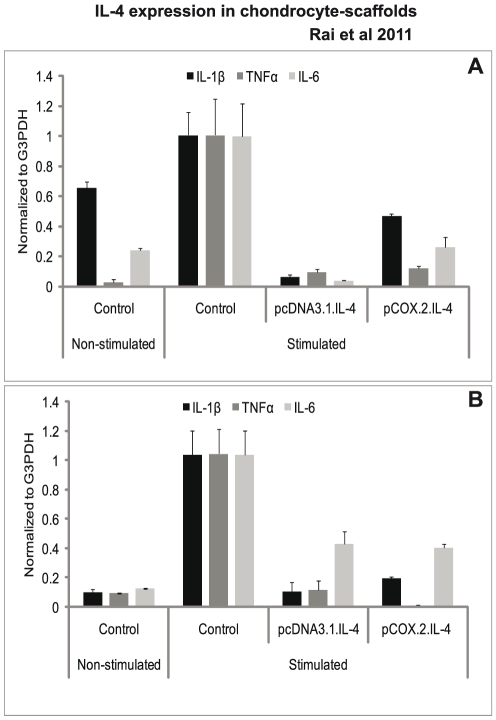
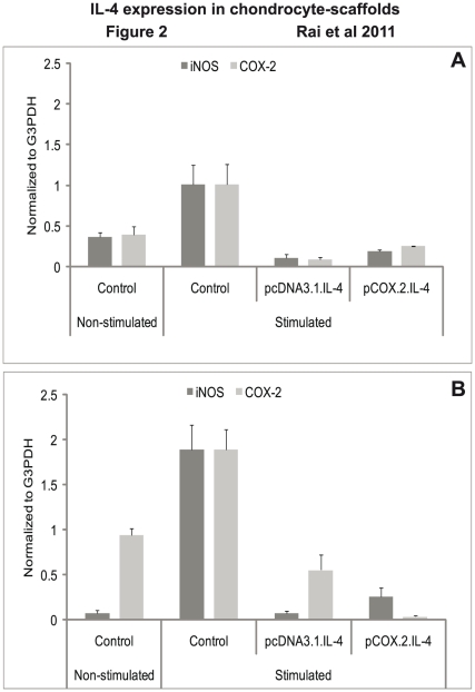
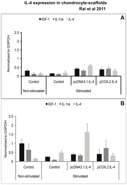
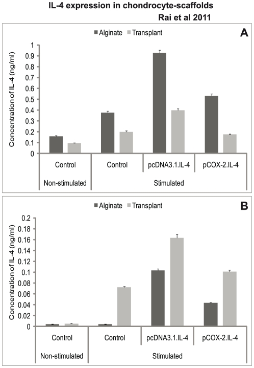
Methods: Mature articular chondrocytes from dogs (n = 3) were conditioned through transient transfection using pcDNA3.1.cIL-4 (constitutive) or pCOX-2.cIL-4 (cytokine-responsive) plasmids. Conditioned cells were seeded in alginate microspheres and rat-tail collagen type I matrix (CaReS®) to generate two types of tissue-engineered 3-dimensional scaffolds. Inflammatory arthritis was simulated in the packed chondrocytes through exogenous addition of recombinant canine (rc) IL-1β (100 ng/ml) plus rcTNFα (50 ng/ml) in culture media for 96 hours. Harvested cells and culture media were analyzed by various assays to monitor the anti-inflammatory and regenerative (anabolic) properties of cIL-4.

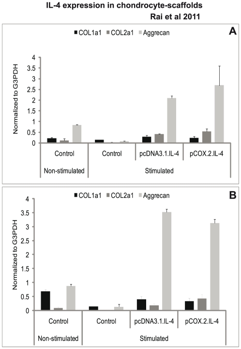
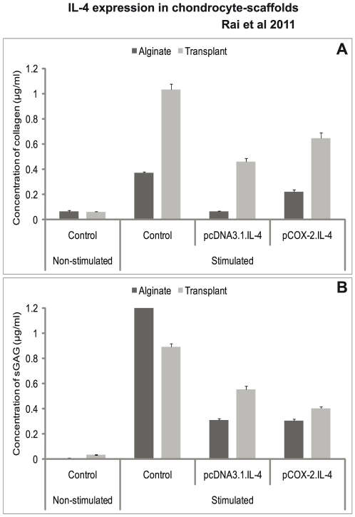
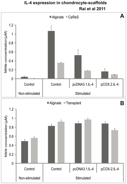
Results: cIL-4 was expressed from COX-2 promoter exclusively on the addition of rcIL-1β and rcTNFα while its expression from CMV promoter was constitutive. The expressed cIL-4 downregulated the mRNA expression of IL-1β, TNFα, IL-6, iNOS and COX-2 in the cells and inhibited the production of NO and PGE(2) in culture media. At the same time, it up-regulated the expression of IGF-1, IL-1ra, COL2a1 and aggrecan in conditioned chondrocytes in both scaffolds along with a diminished release of total collagen and sGAG into the culture media. An increased amount of cIL-4 protein was detected both in chondrocyte cell lysate and in concentrated culture media. Neutralizing anti-cIL-4 antibody assay confirmed that the anti-inflammatory and regenerative effects seen are exclusively driven by cIL-4. There was a restricted expression of IL-4 under COX-2 promoter possibly due to negative feedback loop while it was over-expressed under CMV promoter (undesirable). Furthermore, the anti-inflammatory /anabolic outcomes from both scaffolds were reproducible and the therapeutic effects of cIL-4 were both scaffold- and promoter-independent.

Conclusions: Regulated expression of therapeutic candidate gene(s) coupled with suitable scaffold(s) could potentially serve as a useful tissue-engineering tool to devise future treatment strategies for osteoarthritis.


Conflict of interest statement

Competing Interests: M.F. Rai, S.Twardziok, and M.F.G. Schmidt do not have a conflict of interest in connection with this work. T. Graeve is affiliated with Amedrix GmbH Esslingen Germany. However, his affiliation does not alter the authors' adherence to all the PLoS ONE policies on sharing data and materials, as detailed online in your guide for authors.

Figures

Figure 1
Figure 1. mRNA expression of proinflammatory cytokines.
IL-4 transfected (pcDNA3.1.cIL-4 and pCOX-2.cIL-4) and non-transfected chondrocytes were seeded in alginate microspheres (A) and in CaReS® matrixes (B). Both scaffolds were stimulated with rcIL-1β (100 ng/ml) and rcTNFα (50 ng/ml) for 96 h. mRNA expression was quantified by qRT-PCR. The expression of IL-1, TNF and IL-6 was downregulated in IL-4 expressing scaffolds as compared to non-transfected controls on stimulation.
Figure 2
Figure 2. mRNA expression of enzyme mediators.
IL-4 transfected (pcDNA3.1.cIL-4 and pCOX-2.cIL-4) and non-transfected chondrocytes were seeded in alginate microspheres (A) and in CaReS® matrixes (B). Both scaffolds were stimulated with rcIL-1β (100 ng/ml) and rcTNFα (50 ng/ml) for 96 h. mRNA expression was quantified by qRT-PCR. The expression of iNOS and COX-2 was downregulated in IL-4 expressing scaffolds as compared to non-transfected controls on stimulation.
Figure 3
Figure 3. mRNA expression of regulatory mediators.
IL-4 transfected (pcDNA3.1.cIL-4 and pCOX-2.cIL-4) and non-transfected chondrocytes were seeded in alginate microspheres (A) and in CaReS® matrixes (B). Both scaffolds were stimulated with rcIL-1β (100 ng/ml) and rcTNFα (50 ng/ml) for 96 h. mRNA expression was quantified by qRT-PCR. The expression of IGF-1, IL-1ra and IL-4 was up-regulated in IL-4 expressing scaffolds as compared to non-transfected controls on stimulation.
Figure 4
Figure 4. Detection of IL-4 protein.
IL-4 transfected (pcDNA3.1.cIL-4 and pCOX-2.cIL-4) and non-transfected chondrocytes were seeded in alginate microspheres and in CaReS® matrixes. Both scaffolds were stimulated with rcIL-1β (100 ng/ml) and rcTNFα (50 ng/ml) for 96 h. Cell lysate and culture media were used to measure IL-4 production by sandwich ELISA. A lower IL-4 protein expression was found in cells conditioned with pCOX-2.cIL-4 as compared to those with pcDNA3.1.cIL-4 in lysates (A) and culture media (B). In both constructs, the expression was high as compared to non-transfected chondrocytes.
Figure 5
Figure 5. mRNA expression of collagens and aggrecan.
IL-4 transfected (pcDNA3.1.cIL-4 and pCOX-2.cIL-4) and non-transfected chondrocytes were seeded in alginate microspheres (A) and in CaReS® matrixes (B). Both scaffolds were stimulated with rcIL-1β (100 ng/ml) and rcTNFα (50 ng/ml) for 96 h. mRNA expression was quantified by qRT-PCR. The expression of COL1a1 was downregulated whereas that of COL2a1 and aggrecan was up-regulated in IL-4 expressing scaffolds as compared to non-transfected controls on stimulation.
Figure 6
Figure 6. Release of total collagen and sGAG.
IL-4 transfected (pcDNA3.1.cIL-4 and pCOX-2.cIL-4) and non-transfected chondrocytes were seeded in alginate microspheres and in CaReS® matrixes. Both scaffolds were stimulated with rcIL-1β (100 ng/ml) and rcTNFα (50 ng/ml) for 96 h. Supernatants were used to measure total collagen and sGAG release by respective assays. A diminished release of both collagen (A) and sGAG (B) was observed in the IL-4 expressing scaffolds as compared to non-transfected chondrocytes on stimulation.
Figure 7
Figure 7. Determination of NO production.
IL-4 transfected (pcDNA3.1.cIL-4 and pCOX-2.cIL-4) and non-transfected chondrocytes were seeded in alginate microspheres and in CaReS® matrixes. Both scaffolds were stimulated with rcIL-1β (100 ng/ml) and rcTNFα (50 ng/ml) for 96 h. Culture media were used to measure NO levels by using Griess' reagent system. There was a reduced NO production in IL-4 expressing scaffolds (A). However, there was no reduction in the NO production when the neutralizing anti-cIL-4 antibody was added in the culture media from both scaffolds (B).
Figure 8
Figure 8. PGE2 determination.
IL-4 transfected (pcDNA3.1.cIL-4 and pCOX-2.cIL-4) and non-transfected chondrocytes were seeded in alginate microspheres and in CaReS® matrixes. Both scaffolds were stimulated with rcIL-1β (100 ng/ml) and rcTNFα (50 ng/ml) for 96 h. Culture media were used to measure PGE2 levels by using PGE2 enzyme immunoassay kit. A diminished production of PGE2 was observed in IL-4 expressing scaffolds as compared to non-transfected.

Similar articles

Cited by

References

    1. Pelletier JP, Martel-Pelletier J, Abramson SB. Osteoarthritis, an inflammatory disease: potential implication for the selection of new therapeutic targets. Arthritis Rheum. 2001;44:1237–1247. - PubMed
    1. Hegemann N, Wondimu A, Kohn B, Brunnberg L, Schmidt MF. Cytokine profile in canine immune-mediated polyarthritis and osteoarthritis. Vet Comp Orthop Traumatol. 2005;18:67–72. - PubMed
    1. Nuki G. Role of mechanical factors in the aetiology, pathogenesis and progression of osteoarthritis. In: Reginster JY, Pelletier JP, Martel-Pelletier J, Henrotin Y, editors. Osteoarthritis: Clinical and Experimental Aspects. Berlin: Springer-Verlag; 1999. pp. 101–114.
    1. Pelletier JP, Martel-Pelletier J, Howell DS. Etiopathogenesis of osteoarthritis. In: Koopman WJ, editor. Arthritis & Allied Conditions. A Textbook of Rheumatology. 14th ed. Baltimore: Williams & Wilkins; 2000. pp. 2195–2245.
    1. Sandell LJ, Aigner T. Articular cartilage and changes in arthritis. An introduction: cell biology of osteoarthritis. Arthritis Res. 2001;3:107–113. - PMC - PubMed

Publication types

MeSH terms