

T-helper cell-mediated proliferation and cytokine responses against recombinant Merkel cell polyomavirus-like particles
- PMID: 21991346
- PMCID: PMC3185038
- DOI: 10.1371/journal.pone.0025751
T-helper cell-mediated proliferation and cytokine responses against recombinant Merkel cell polyomavirus-like particles
Abstract
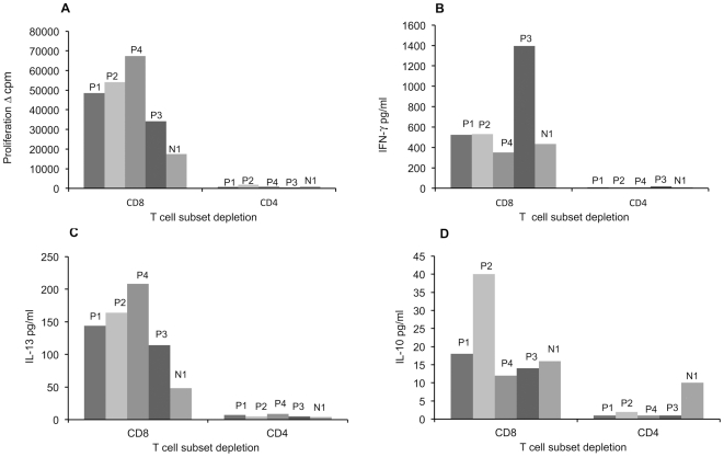
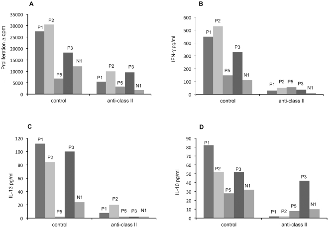
The newly discovered Merkel Cell Polyomavirus (MCPyV) resides in approximately 80% of Merkel cell carcinomas (MCC). Causal role of MCPyV for this rare and aggressive skin cancer is suggested by monoclonal integration and truncation of large T (LT) viral antigen in MCC cells. The mutated MCPyV has recently been found in highly purified leukemic cells from patients with chronic lymphocytic leukemia (CLL), suggesting a pathogenic role also in CLL. About 50-80% of adults display MCPyV-specific antibodies. The humoral immunity does not protect against the development of MCC, as neutralizing MCPyV antibodies occur in higher levels among MCC patients than healthy controls. Impaired T-cell immunity has been linked with aggressive MCC behavior. Therefore, cellular immunity appears to be important in MCPyV infection surveillance. In order to elucidate the role of MCPyV-specific Th-cell immunity, peripheral blood mononuclear cells (PBMC) of healthy adults were stimulated with MCPyV VP1 virus-like particles (VLPs), using human bocavirus (HBoV) VLPs and Candida albicans antigen as positive controls. Proliferation, IFN-γ, IL-13 and IL-10 responses were examined in 15 MCPyV-seropositive and 15 seronegative volunteers. With the MCPyV antigen, significantly stronger Th-cell responses were found in MCPyV-seropositive than MCPyV-seronegative subjects, whereas with the control antigens, the responses were statistically similar. The most readily detectable cytokine was IFN-γ. The MCPyV antigen tended to induce stronger IFN-γ responses than HBoV VLP antigen. Taken together, MCPyV-specific Th-cells elicit vigorous IFN-γ responses. IFN-γ being a cytokine with major antiviral and tumor suppressing functions, Th-cells are suggested to be important mediators of MCPyV-specific immune surveillance.
Conflict of interest statement
Figures
References
-
- Kassem A, Schopflin A, Diaz C, Weyers W, Stickeler E, et al. Frequent detection of Merkel cell polyomavirus in human Merkel cell carcinomas and identification of a unique deletion in the VP1 gene. Cancer Res. 2008;68:5009–5013. - PubMed
-
- Andres C, Belloni B, Puchta U, Sander CA, Flaig MJ. Prevalence of MCPyV in Merkel cell carcinoma and non-MCC tumors. J Cutan Pathol. 2010;37:28–34. - PubMed
-
- Becker JC, Houben R, Ugurel S, Trefzer U, Pfohler C, et al. MC polyomavirus is frequently present in Merkel cell carcinoma of european patients. J Invest Dermatol. 2009;129:248–250. - PubMed
Publication types
MeSH terms
Substances
LinkOut - more resources
Full Text Sources
