. 2011;6(10):e25900.
doi: 10.1371/journal.pone.0025900. Epub 2011 Oct 4.

Sclerostin stimulates osteocyte support of osteoclast activity by a RANKL-dependent pathway

Affiliations

Sclerostin stimulates osteocyte support of osteoclast activity by a RANKL-dependent pathway

Asiri R Wijenayaka et al. PLoS One. 2011.

Abstract

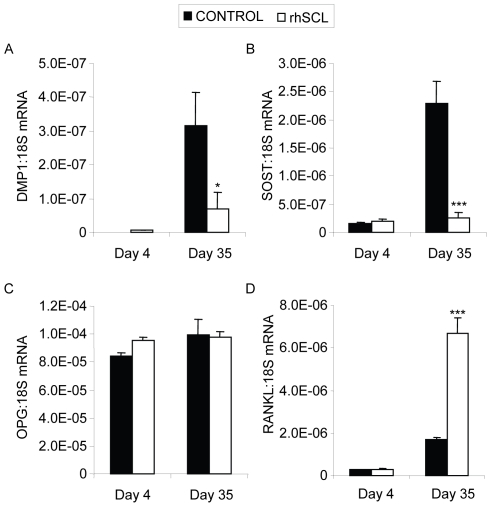
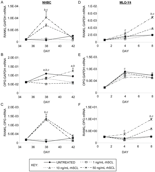
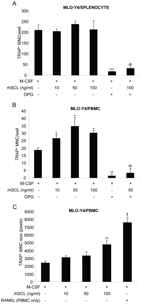
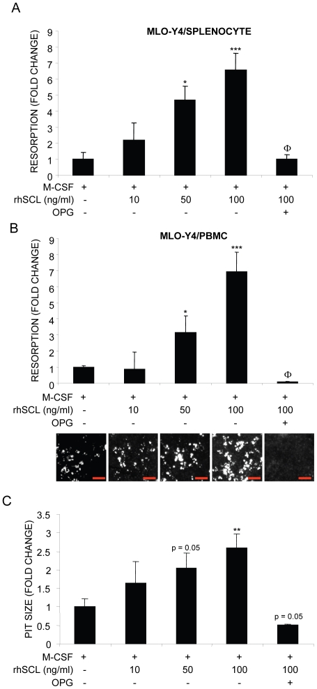
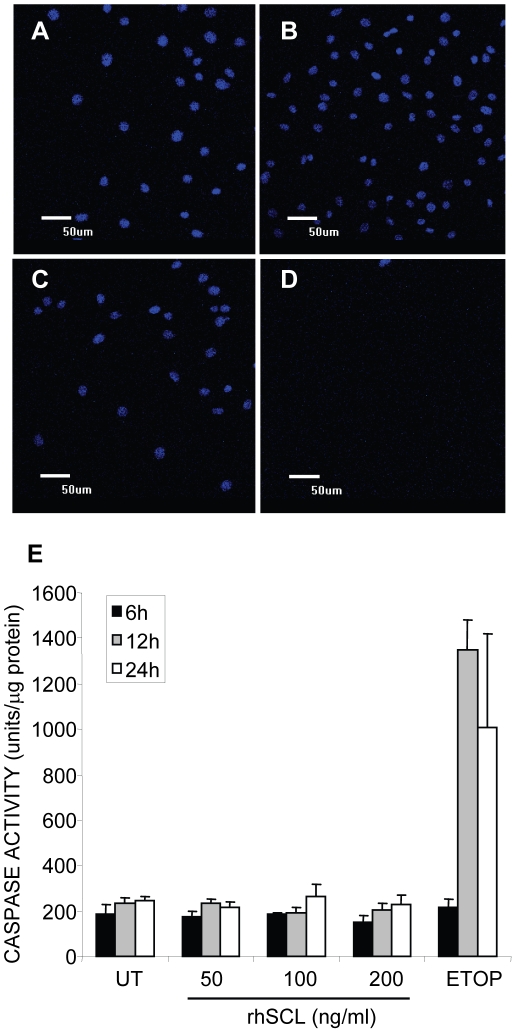
Sclerostin is a product of mature osteocytes embedded in mineralised bone and is a negative regulator of bone mass and osteoblast differentiation. While evidence suggests that sclerostin has an anti-anabolic role, the possibility also exists that sclerostin has catabolic activity. To test this we treated human primary pre-osteocyte cultures, cells we have found are exquisitely sensitive to sclerostin, or mouse osteocyte-like MLO-Y4 cells, with recombinant human sclerostin (rhSCL) and measured effects on pro-catabolic gene expression. Sclerostin dose-dependently up-regulated the expression of receptor activator of nuclear factor kappa B (RANKL) mRNA and down-regulated that of osteoprotegerin (OPG) mRNA, causing an increase in the RANK:OPG mRNA ratio. To examine the effects of rhSCL on resulting osteoclastic activity, MLO-Y4 cells plated onto a bone-like substrate were primed with rhSCL for 3 days and then either mouse splenocytes or human peripheral blood mononuclear cells (PBMC) were added. This resulted in cultures with elevated osteoclastic resorption (approximately 7-fold) compared to untreated co-cultures. The increased resorption was abolished by co-addition of recombinant OPG. In co-cultures of MLO-Y4 cells with PBMC, SCL also increased the number and size of the TRAP-positive multinucleated cells formed. Importantly, rhSCL had no effect on TRAP-positive cell formation from monocultures of either splenocytes or PBMC. Further, rhSCL did not induce apoptosis of MLO-Y4 cells, as determined by caspase activity assays, demonstrating that the osteoclastic response was not driven by dying osteocytes. Together, these results suggest that sclerostin may have a catabolic action through promotion of osteoclast formation and activity by osteocytes, in a RANKL-dependent manner.


Conflict of interest statement

Competing Interests: The authors have declared that no competing interests exist.

Figures

Figure 1
Figure 1. Effects on gene expression of continuous exposure of mineralizing NHBC cultures to rhSCL.
NHBC were cultured under mineralizing conditions for up to 35 days in the absence or presence of rhSCL at 50 ng/ml. Media were replenished every 3–4 days. At the time points indicated, total RNA was prepared and real-time RT-PCR performed to determine mRNA expression of A) DMP1, B) SOST, C) OPG and D) RANKL. Data shown are means of triplicate reactions ± SD normalized to expression of 18S mRNA. Significant differences to untreated control are indicated by * p<0.05 and *** p<0.001. Similar results were obtained from 3 independent experiments using NHBC from different donors.
Figure 2
Figure 2. Effects on gene expression of acute exposure of osteocyte-like cells to rhSCL.
Human osteocyte-like cultures, derived from NHBC cultured under mineralizing conditions for 35 days, or cultures of MLO-Y4 cells were cultured for 3 or 7 days in the absence of presence of rhSCL at 1, 10 or 50 ng/ml. Media and supplements were replenished at day 3. Total RNA was prepared and real-time RT-PCR performed to determine mRNA expression of A) human RANKL, B) human OPG or mouse Rankl (D) and Opg (E). The ratios of RANKL∶OPG mRNA for NHBC (C) and MLO-Y4 (D) were also determined. Data shown are means of triplicate reactions ± SD normalized to expression of GAPDH mRNA. a, b and c indicate significant differences (p<0.05) to untreated control for rhSCL at 1, 10 and 50 ng/ml, respectively. Near identical results were obtained from 3 independent experiments using NHBC from different donors or MLO-Y4 cells.
Figure 3
Figure 3. Effects of rhSCL on TRAP+ multinucleated cell formation.
The effect of rhSCL on osteoclastogenesis was tested in co-cultures of MLO-Y4 cells with A) mouse splenocytes and B) human PBMC. In both cases MLO-Y4 cells were seeded into type I collagen coated culture wells and cultured for 72 h in the absence or presence of rhSCL, as indicated, prior to the addition of either splenocytes or PBMC. All cultures received rhM-CSF at 25 ng/ml. RhOPG was added to some cultures at 100 ng/ml and rhRANKL (100 ng/ml) added to monocultures of either splenocytes or PBMC to confirm the osteoclast-forming potential of these populations (not shown). Media were replenished every 3 days. Cultures were fixed and stained for TRAP at day 6. TRAP-positive MNC, defined as cells with >3 nuclei, were counted from quadruplicate wells. Asterisks denote significant differences to the no SCL control (** p<0.01, *** p<0.001) and the effect of OPG relative to the corresponding SCL-only treatment is indicated by Φ (p<0.001). C) In the case of MLO-Y4/PBMC co-cultures and cells formed in PBMC monocultures treated with rhRANKL, the relative size (in pixels) of TRAP+ MNC formed (in each case >50 cells) was measured by Image J analysis. Difference to the no SCL control is indicated by ** p<0.01; † indicates difference of rhRANKL control to rhSCL at 100 ng/ml (p<0.01). Data shown are representative of at least 3 independent experiments.
Figure 4
Figure 4. Effects of rhSCL on osteoclast resorptive activity.
Co-cultures of MLO-Y4 cells and A) mouse splenocytes and B) human PBMC were established, in which MLO-Y4 cells were seeded onto bone-like Osteologic™ slides and cultured for 72 h in the absence or presence of rhSCL as indicated prior to the addition of either splenocytes or PBMC. All cultures received rhM-CSF at 25 ng/ml and some cultures were treated with rhOPG (100 ng/ml). Media were replenished every 3 days for 14 days. Cells were removed and slides developed as described in Materials and Methods and resorption. The entire surface areas of the developed slides were imaged using an Olympus SZX10 dissecting microscope at high resolution, depicted for the PBMC co-cultures below the corresponding histograms in panel B) (red bars indicate 500 µm). Total resorbed area was then quantified from quadruplicate wells using ImageJ software. C) In the case of MLO-Y4/PBMC co-cultures, mean resorption pit size was determined and expressed as fold-change from the corresponding untreated co-culture. Differences to the ‘no SCL’ control in each panel are indicated by * p<0.05, ** p<0.01, *** p<0.001. The effect of OPG relative to the corresponding SCL-only treatment is indicated by Φ (p<0.001). Data shown are representative of 3 independent experiments.
Figure 5
Figure 5. Effect of rhSCL on MLO-Y4 viability.
A) Cells were seeded into chamber slides and treated for 3 days A) untreated, or in the presence of rhSCL at 50 ng/ml (B) or 100 ng/ml (C), or D) in the presence of etoposide (50 µM). Cells were then fixed and their nuclear morphology determined by staining with DAPI and visualization by confocal microscopy. E) Cells were seeded into wells of a 6-well plate and cultured for the times indicated with rhSCL (0–200 ng/ml) or in the presence of etoposide (ETOP) at 50 µM. After the times indicated, cell lysates were prepared and caspase 3 activity determined, as described in Materials and Methods. Data are expressed as means of quadruplicate wells ± standard deviations.

References

    1. Baron R, Rawadi G, Roman-Roman S. Wnt signaling: a key regulator of bone mass. Curr Top Dev Biol. 2006;76:103–127. - PubMed
    1. Li X, Ominsky MS, Niu QT, Sun N, Daugherty B, et al. Targeted Deletion of the Sclerostin Gene in Mice Results in Increased Bone Formation and Bone Strength. J Bone Miner Res. 2008;23:860–869. - PubMed
    1. ten Dijke P, Krause C, de Gorter DJ, Lowik CW, van Bezooijen RL. Osteocyte-derived sclerostin inhibits bone formation: its role in bone morphogenetic protein and Wnt signaling. J Bone Joint Surg Am. 2008;90(Suppl 1):31–35. - PubMed
    1. Balemans W, Patel N, Ebeling M, Van Hul E, Wuyts W, et al. Identification of a 52 kb deletion downstream of the SOST gene in patients with van Buchem disease. J Med Genet. 2002;39:91–97. - PMC - PubMed
    1. Staehling-Hampton K, Proll S, Paeper BW, Zhao L, Charmley P, et al. A 52-kb deletion in the SOST-MEOX1 intergenic region on 17q12-q21 is associated with van Buchem disease in the Dutch population. Am J Med Genet. 2002;110:144–152. - PubMed

Publication types

MeSH terms